
Jovem masculino, Dispneia de repouso iniciada um mês após COVID
Mês: maio 2021

Microfunção ao ecocardiograma

https://www.frontiersin.org/articles/10.3389/fcvm.2021.670734/full
Discussion
In this pilot study we demonstrate a unique multiscale, multimodality protocol to visualize in vivo and ex vivo myocardial tissue with non-invasive and X-PCI imaging. We report a novel approach to visualize 3D cardiac microstructure in LVH and relate the findings to the etiology-discriminative interpretation of non-invasive imaging data.
3D não é só válvula Mitral!!!
Agora é realidade
Ecoestresse 3D funciona
Você escolhe se quer tranquilidade e conforto fazendo o 2D apical 3 em 1 para 4, 2 e 3 Câmaras
Ou usa o 3D “fatia” e coleta as janelas com a profundidade do 3D
Ou faz a escolha depois de gravar tudo!
Tantas opções, encapuzadas na rotina de um laboratório onde o tempo é tridimensional
Mais um mito detonado: Aparelho 3D não serve para a rotina!

EchoTalk desfaz mais um mito da ecocardiografia.
Existia o mito que o 3D não traria benefícios para um laboratório de ecocardiografia privado
Provamos que o E9 pode acelerar os exames Ecoestresse e ainda poupar seu ombro.
Com ganhos no tempo e na análise do exame

Pensando em ajudar com Carótidas e Ecoestresse
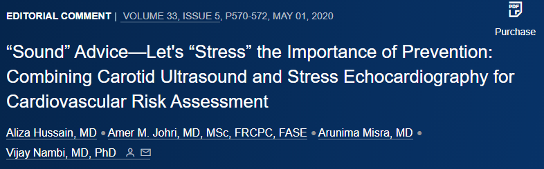
https://doi.org/10.1016/j.echo.2020.03.003
‘‘Sound’’ Advice—Let’s ‘‘Stress’’ the
Importance of Prevention: Combining
Carotid Ultrasound and Stress
Echocardiography for Cardiovascular Risk
Assessment
In conclusion, assessment of plaque with a carotid US as a supplement to functional SE provides incremental improvement in risk prediction and can prove clinically useful (especially in patients with a
normal SE) to guide primary prevention. However, practical issues
related to exact timing of US and SE, training of echocardiographers,
and cost-effectiveness and coverage of the combined modalities will
be important determinants of whether this approach is adopted clinically for cardiovascular risk assessment.
Aplicação do 3D no Ecoestresse
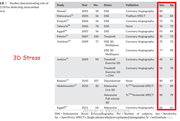
REVIEW
Stress echocardiography using real-time three-dimensional imaging
Rafic F. Berbarie MD, Elie Dib MD, Masood Ahmad MD, FACC, FASE
Division of Cardiology, Department of Internal Medicine, University of Texas Medical Branch, Galveston, TX, USA
With its ability to acquire the entire LV volume in a single beat,
RT-3D-SE offers substantial promise for becoming the reference
standard in the evaluation of patients with suspected CAD.
.
Estamos aplicando o futuro hoje, Quarta, dia 26. Ano 2021.
Aorta para o corpo

https://doi.org/10.1161/CIRCIMAGING.116.005121
Conclusions—
Among AVA normalization methods, AVA/height <0.45 cm2/m followed by AVA/BSA <0.40 cm2/m2 seem as robust parameters for defining high risk in asymptomatic aortic stenosis. The prognostic value of AVA/height deserves future research.