

.
Novos nomes e métodos antigos, essa é a rotina da prova do título de ecocardiografista.
.
Prova que testa mémoria, não conhecimento da técnica. Questões sobre métodos que são usados por 5% dos ecocardiografistas brasileiros…
.
Prova longa, perguntas de demorada interpretação, algumas confusas…
.
Seguimos barrando ecocardiografistas bons, elitizando um método que precisa se popularizar para sobreviver!
.
Quem vai ter coragem de mudar essa prova?
.
Mês: maio 2010
Quando o Ateroma é móvel, é preciso informar.

.
Estudo mostra a forte correlação do ateroma com mobilidade e eventos embólicos.
.
A pesquisa de ateroma da aorta, relegada a um segundo plano, sempre foi importante clinicamente.
.
Nem tudo que NÃO brilha, é colesterol.
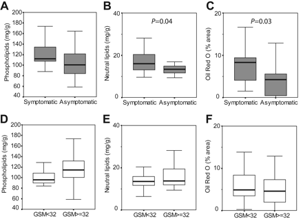
Results— As assessed biochemically (milligrams per gram [mg/g]), echolucent plaques contained less hydroxyapatite (43.8 [SD 41.2] mg/g versus 121.6 [SD 106.2] mg/g; P=0.018), more total elastin (1.7 [SD 0.4] mg/g versus 1.2 [SD 0.4] mg/g; P=0.008), and more intermediate-size elastin forms (1.2 [SD 0.3] mg/g versus 0.8 [SD 0.4] mg/g; P=0.018). There was no difference in collagen amount between echogenic and echolucent plaques, neither biochemically (15.3 [SD 3.7] mg/g versus 14.4 [SD 3.4] mg/g) nor histologically (13.4 [SD 4.9] % versus 13.0 [SD 5.6] %). Cholesterol esters, unesterified cholesterol, and triglycerides were increased in plaques associated with symptoms (22.5 [SD 23.3] mg/g versus 13.3 [SD 3.2]; P=0.04), but no differences were detected between echolucent and echogenic plaques (13.5 [SD 4.0] versus 20.2 [SD 21.5] mg/g). Similar results were obtained by Oil Red O staining (symptomatic 7.6 [SD 4.7] % versus asymptomatic 4.2 [SD 3.6] %; P=0.03; echolucent 5.9 [SD 4.1] % versus echogenic 5.0 [SD 4.0] % of area).
.
Conclusions— Echogenicity of carotid plaques is mainly determined by their elastin and calcium but not collagen or lipid content. In addition, echolucency is associated to higher elastin content.
Stroke. 2004;35:2795
.
Pesquisa muito boa, mostrando que a ecolucência ( Escuro ao ultrassom) se relaciona mais a presença de elastina do que colesterol nas placas de carótida!
.
Então quando a placa parece escura após a capa fibrosa, não é possível afirmar que é lípide?
.
Isso muda muitos conceitos…
.
Fibrilação atrial e Strain do átrio esquerdo

Aqui
.
Left Atrial Strain and Strain Rate in Patients With Paroxysmal and Persistent Atrial Fibrillation
.
Circulation: Cardiovascular Imaging. 2010;3:231-239
.
Conclusion, LA wall fibrosis, implied by delayed enhancement on MRI, is inversely related to LA strain and strain rate. Echocardiographic assessment of structural and functional remodeling of the LA is possible from gray-scale images. These techniques appear to have some discriminatory power in assessing patients with paroxysmal versus persistent AF. Combined, noninvasive imaging with atrial strain and strain rate and quantification of LA wall fibrosis by DE-MRI may be helpful in predicting outcomes and guiding therapeutic strategies in patients with AF. Novel imaging strategies such as those described in this report may allow us to identify patients earlier in their disease process, before the development of severe or irreversible abnormalities. Echocardiographic strain imaging of the atria may be particularly useful in centers where DE-MRI is unavailable or in patients who are not candidates for MRI.
.
Ao final do curso preparatório pra prova, um aluno me perguntou qual das novas tecnologias parecia mais promissora.
Respondi de pronto: Strain com uso do Speckle Traking.
.
No artigo acima, a análise do Strain no Atrio esquerdo ajuda a entender o grau de doença já presente no átrio e com isso, as chances de reversão permanente da fibrilação.
Função do Ventrículo esquerdo: novo modo, fácil e rápido.

Rapid Estimation of Left Ventricular Function Using Echocardiographic Speckle-Tracking of Mitral Annular Displacement
.
Journal of the American Society of Echocardiography
Issue: Volume 23(5), May 2010, p 511–515
.
Conclusions: STE MAD is a clinically useful tool for quick, easy, robust, and accurate estimates of EF irrespective of LV endocardial definition.
.
Mais fácil que fazer o Simpson, se o ECG estiver ligado, o movimento do anel mitral captado pelo Speckle-Tracking pode ser usado como medida da função ventricular.
Desde o modo M se tenta usar o movimento do anel mitral como referência para a função sistólica, o que faz muito sentido pois a contração é predominantemente longitudinal ao repouso.
.
Diretamente de Chicago, uma técnica simples, rápida e útil.
.
3D, um exame com futuro, no futuro.
.
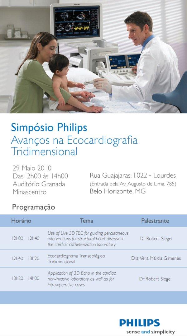
Há 10 anos sou convidado para aulas sobre o avanço do 3D.
Nesse tempo, a internet saiu do modem e virou 3G.
A China virou potência mundial e fabrica carros e computadores.
Celular, mini-notebooks, tomo de coronária, eco portátil, mp3, bluray…
.
E o 3D continua sendo para amanhã…
.
Miocárdio não compactado revertido(?)

.
We conclude that diagnostic features of IVNC based on widely used echocardiographic criteria may be apparent in left ventricular systolic dysfunction due to other causes. Although the diagnosis should be considered at an early stage, caution should be exercised in making a diagnosis of IVNC, particularly during an acute presentation. Follow-up imaging should be performed to look for possible resolution of IVNC features, which may suggest an alternative diagnosis.
.
Revista
destaca caso listado como não compactação das fibras do miocárdio que reverteu após tratamento da ICC.
.
Com a explosão do número de diagnósticos, vários casos que eram apenas insuficiência cardíaca, foram catalogados como miocárdio não compactado.
.
Veja na foto acima um típico caso, com os achados necessários para um diagnóstico tão sombrio.
.