
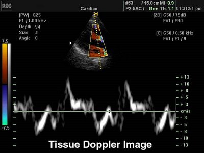
.
Na medicina, devemos aliviar os sintomas e prolongar a vida.
Sem prolongar, é obrigatório aliviar os sintomas.
Os sintomas principais da insuficiência cardíaca se dão pelo aumento da pressão no átrio esquerdo, transmitida aos capilares pulmonares.
Então, estimar a pressão no átrio esquerdo é fundamental!
.
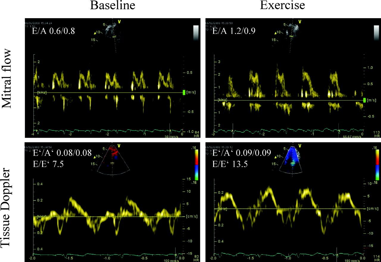
A procura de um marcador único, como o E/e´, revela um pensamento limitado.
Toda análise ao ecocardiograma deve incluir múltiplos parâmetros.
Para a análise da diástole, serão analisados:
Volume do átrio esquerdo.
Onda E, Onda A.
Tempo de despressurização atrial
Tempo de relaxamento isovolumétrico.
Onda e´, Onda a´. Septal e lateral.
Velocidade de propagação.
Fluxo de veias pulmonares.
Volume ventricular.
Relação E/e´.
Faltou algum?
Pelo menos 5 marcadores para uma resposta adequada!
Dia: 9 de setembro de 2009
A avaliação diastólica não ajuda o paciente?
Doppler Echocardiography Yields Dubious Estimates of Left Ventricular Diastolic Pressures
.Circulation. 2009;120:810-820
.
Doppler echocardiography provided major insights into the pathophysiology of diastolic LV dysfunction. So far, however, no single Doppler echocardiographic index of diastolic LV dysfunction has yielded a robust criterion for elevated LV filling pressures. Furthermore, a stepwise strategy with sequential use of multiple Doppler echocardiographic indexes reduces diagnostic sensitivity because it frequently leads to an indeterminate outcome with a positive score on 1 test and a negative on another
Because of these persistent shortcomings, clinicians should continue to make critical use of current Doppler echocardiographic estimates of LV filling pressures and should not hesitate to implement invasive investigations to confirm their clinical suspicions.
A avaliação diastólica ajuda o paciente?

Is echocardiographic evaluation of diastolic function useful in determining clinical care?
Circulation. 2009;120:802-809
.
An increased E wave in the presence of a reduced e’ (ie, E/e’ >15) indicates elevated LA pressure. In patients with borderline increased E/e’ (>8, <15), the entire echocardiographic picture should be assessed, including LA size, mitral filling pattern, DT, pulmonary vein velocities, and pulmonary artery systolic pressure. This information is particularly helpful in establishing the diagnosis in patients with the clinical picture of heart failure when the LV EF is preserved. It provides prognostic information and can help guide therapy in patients with heart failure regardless of the EF.