
.
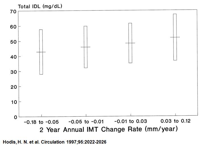
In conclusion, our data present compelling evidence that lipoproteins in the IDL fraction (Sf 12 to 20) are associated with progression of atherosclerosis as directly quantified by arterial wall IMT measurements, whereas lipoproteins in the LDL and VLDL density ranges are not associated with progression of carotid IMT.
.
Tradicionalmente, quando medimos o LDL, dosamos junto com o IDL.
Estudos que mostraram benefícios clínicos das estatinas, a maioria deles, usou o LDL “misturado” com o IDL.
Este estudo ressalta a importância das moléculas ricas em triglicérides na gênese da Aterosclerose acelerada.
Têm a ver com os triglicérides a aceleradíssima Aterosclerose dos Diabéticos?
Em meu estudo pós cirurgia bariátrica, a espessura da carótida despencou junto com os triglicérides…
.
Belo artigo, que só li agora…
(So many things, so little time)
.
Mês: julho 2010
Dissecção da aorta no Circulation

Clinician Update
Alan C. Braverman, MD
.
The selection of a specific imaging modality is influenced by individual patient characteristics and variables and institutional capabilities and expertise. Contrast computed tomography is the most commonly used modality in diagnosing aortic dissection and is readily available in most emergency rooms. Because magnetic resonance imaging takes longer for image acquisition and leaves the patient relatively unmonitored, it is usually not the procedure of first choice. Transesophageal echocardiogram has an advantage in being portable and able to be performed at the bedside for the unstable patient. Although aortic dissection may be diagnosed by transthoracic echocardiogram, the sensitivity and specificity are much lower than with the other diagnostic modalities; thus, this technique is not the first choice for diagnosing dissection.
.
Realmente o ecocardiograma transtorácico não é a modalidade de escolha e pode gerar uma falsa segurança ao clínico.
Analisando o quadro clínico podemos pensar:
– Toda suspeita considerável de dissecção da aorta deve ser colocada em repouso absoluto, deve receber analgesia com opiáceo e receber drogas em bomba de infusão para controlar o duplo produto.
.
Quer paciente mais fácil e apropriado para o exame transesofágico?!?!
.
Sem contraste, sem demora, sem x vezes a gravidade da terra!
.
Sensibilidade 98% Especificidade 100%.
Custo inferior a qualquer tomo ou ressonância.
.
Ou não é uma suspeita de Dissecção a ser levada a sério(Vai para Tomo…), ou têm que fazer transesofágico.
.
Todo exame deve ir aonde o paciente está.

Tissue Doppler echocardiography reveals distinct patterns of impaired myocardial velocities in different degrees of coronary artery disease
.
Aqui
.
Methods and results This study comprises 82 patients with suspected angina pectoris, no previous cardiac history, and a normal ejection fraction, who were all examined with colour TDI prior to coronary angiography. Patients without significant stenoses (n = 35) constituted the control group and patients with significant stenoses (n = 47) were divided into three groups according to significant one-, two-, or three-vessel disease (n = 18, n = 14, and n = 15, respectively). Regional longitudinal peak systolic (s′), early (e′), and late diastolic (a′) myocardial velocities were measured at six mitral annular sites and averaged to provide global estimates. Each patient with significant coronary disease was matched with a control of the same age, sex, body mass index, and status regarding diabetes and hypertension. Global systolic and diastolic performance by TDI (in terms of global s′ and E/e′) were negatively correlated to the number of vessels with significant stenoses (both P < 0.05). Regional analyses revealed that in one- and two-vessel disease, e′ decreased significantly in the segments supplied by a stenotic artery. In patients with one-vessel disease, a′ increased compensatorily with a significant reduction of e′/a′-ratio (0.86 ± 0.24 vs. 1.00 ± 0.28, P < 0.05). Both regional and global s′ was significantly reduced in patients with three-vessels disease.
Conclusion Colour TDI performed at rest in patients with stable angina and preserved ejection fraction reveals both diastolic and systolic dysfunction and the nature of the dysfunction depends on the extent of the CAD.
.
Detectar isquemia ao repouso, em um ecocardiograma, sempre foi o sonho dos ecocardiografistas.
Esse estudo levanta a possibilidade de detectar estenoses com o Doppler tecidual, diferenciando “uni” de “bi” e triarterial.
.
Imagine essa técnica em um pronto atendimento, usando um portátil bom nas mãos de um médico adequadamente treinado!
Custo? 100 vezes menor que a Tomo ou Cintilografia(no total de investimento)
Radiação e contraste? Zero.
Possibilidade de repetição do exame no mesmo dia? Quantas vezes forem necessárias.
.
O problema da dor torácica sem diagnóstico no PA pode ser resolvido pela ecocardiografia.
.
Olha o Eco de esforço aí, gente!

Exercise Pulmonary Hypertension in Asymptomatic Degenerative Mitral Regurgitation
.
Circulation. 2010;122:33-41
.
Methods and Results— Comprehensive resting and exercise transthoracic echocardiography was performed in 78 consecutive patients (age, 61±13 years; 56% men) with at least moderate degenerative mitral regurgitation (effective regurgitant orifice area =43±20 mm2; regurgitant volume =71±27 mL). Exercise PHT was defined as a systolic pulmonary arterial pressure (SPAP) >60 mm Hg. Exercise PHT was present in 46% patients. In multivariable analysis, exercise effective regurgitant orifice was an independent determinant of exercise SPAP (P<0.0001) and exercise PHT (P=0.002). Resting PHT and exercise PHT were associated with markedly reduced 2-year symptom-free survival (36±14% versus 59±7%, P=0.04; 35±8% versus 75±7%, P56 mm Hg) was more accurate than resting PHT (SPAP >36 mm Hg) in predicting the occurrence of symptoms during follow-up (P=0.032).
Conclusions— Exercise PHT is frequent in patients with asymptomatic degenerative mitral regurgitation. Exercise mitral regurgitation severity is a strong independent predictor of both exercise SPAP and exercise PHT. Exercise PHT is associated with markedly low 2-year symptom-free survival, emphasizing the use of exercise echocardiography. An exercise SPAP >56 mm Hg accurately predicts the occurrence of symptoms.
.
Para melhorar nossa semana, vejam artigo que comprova mais uma utilidade do ecostress na bicicleta.
.
Essa história de ficar esperando grandes sintomas para intervir em válvulas um dia vai acabar…
.
Olha o Strain aí, gente!
.
ao vivo
.
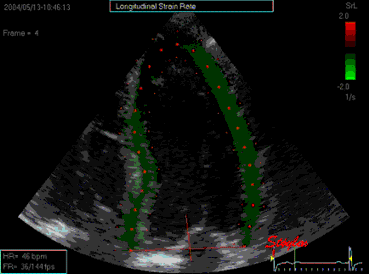
Strain echocardiography predicts acute coronary occlusion in patients with non-ST-segment elevation acute coronary syndrome
.
An area of ≥4 adjacent dysfunctional segments (strain greater than or equal to −14%) had the best ability to identify patients with acute coronary occlusion, with sensitivity 85% and specificity 70%. WMS demonstrated slightly less accuracy than strain.
.
Conclusion Strain echocardiography identifies NSTE-ACS patients with acute coronary occlusion, who may benefit from urgent reperfusion therapy.
.
O Strain longitudinal ganha força todo dia!
.

Embolização e o Ecocardiograma

.
Recommendations for echocardiography use in the diagnosis and management of cardiac sources of embolism
.
Table 1
Potential cardioembolic sources
Major risk sources
Atrial fibrillation
Recent myocardial infarction
Previous myocardial infarction (LV aneurysm)
Cardiomyopathies
Cardiac masses
Intracardiac thrombus Atrial septal aneurysm
Intracardiac tumours
Fibroelastoma Patent foramen ovale
Marantic vegetations
Rheumatic valve disease (mitral stenosis)
Aortic arch atheromatous plaques
Endocarditis
Mechanical valve prosthesis
.
Table 3
Echocardiographic predictors of embolic risk in patients with atrial fibrillation
Echocardiographic risk factors
Left ventricular systolic dysfunction (EF < 35%)
Complex aortic plaquesa
LAA thrombi or spontaneous echo contrasta
LAA dysfunction (emptying blood flow velocities ≤20 cm/s and/or reduced contraction at M-mode)
.
Conclusion:
A potential cardiac source of embolism should be considered in all patients presenting with stroke or TIA. In this regard, echocardiography is not only a powerful tool for the evaluation of cardioembolic sources of stroke, but also to establish recommendations for the primary and secondary prevention of cardioembolic stroke.