Ecostress em mulheres

Exercise Stress Testing in Women

Going Back to the Basics

.
Both exercise stress echocardiography and SPECT techniques have limitations that are specific to women and must be taken into consideration. In exercise SPECT imaging, a false-positive test result can occur because of soft tissue attenuation due to either breast tissue or body habitus. Other gender differences in exercise SPECT may be explained by reduced left ventricular cavity size and smaller coronary arteries in women. Stress echocardiography may be limited by the variability in acoustic windows and the ability to capture images at the point of maximal stress. Despite these limitations, both the diagnostic accuracy and prognostic value of stress SPECT and stress echocardiography exceed those of exercise ECG alone, with no significant differences between men and women.
.
Diagnostic Value
Exercise stress echocardiography has a higher specificity and sensitivity than exercise ECG alone, increasing the specificity and sensitivity to 81% to 86% and 80% to 88%, respectively, for diagnosis of obstructive coronary disease in symptomatic women.
There has been no direct comparison of the diagnostic ability of exercise SPECT and exercise echocardiography. In a systematic review of the literature of exercise testing with imaging that included women, the results of 10 studies in which SPECT was used and 4 studies in which echocardiography was used were combined. There was no difference between exercise SPECT and exercise echocardiography in the sensitivity (77% and 81%, respectively) and specificity (63% and 73%, respectively) for the diagnosis of CAD in women.
.
Although these imaging modalities have not been compared directly, it appears that exercise stress echocardiography and SPECT have similar prognostic value in the prediction of cardiac events and death in women. In a meta-analysis of exercise echocardiography and SPECT imaging by Metz et al, both exercise imaging modalities had the same NPV in women (99% versus 98%, respectively), which was similar to our findings in men. The authors suggested that the use of either imaging modality was appropriate for either gender, and the choice of test should depend on the experience and cost at individual institutions.
.
Bom, é isso.
Use o tradutor acima se tiver dificuldades com o Inglês.
Mas o assunto está encerrado.
São iguais e um deles é mais barato e rápido!
.


A avaliação funcional das estenoses é a melhor opção e se paga.

Economic Evaluation of Fractional Flow Reserve–Guided Percutaneous Coronary Intervention in Patients With Multivessel Disease

Circulation. 2010;122:2545-2550
.

The principal finding of this study is that performing PCI guided by FFR in patients with multivessel CAD saves healthcare resources and improves health outcomes at 1 year compared with a traditional strategy of angiographic guidance. 
.
The presumed explanation for these results lies in the fact that the coronary angiogram, although our reference standard for diagnosing CAD, is a poor predictor of ischemia-producing lesions. For example, in the FAME trial, 65% of lesions with 50% to 70% diameter stenosis based on visual estimation were not responsible for an abnormal FFR (≤0.80), and 20% of lesions with a 71% to 90% diameter stenosis were not responsible for an abnormal FFR
.
.
Mesmo o óbvio precisa ser provado.
Todos sabíamos que colocar stents em lesões que não provocam isquemia era desperdício de dinheiro.
.
Todos sabíamos, após o US coronário, que o cateterismo era um método irregular de avaliar o potencial para isquemia das  estenoses.
Descobrimos que um Cateterismo normal poderia corresponder a até 20% de lesoões ao US.
.
O estudo acima comprovou o que o Ecostress faz há mais de 30 anos.
Só quando tem isquêmia demonstrada vale a pena intervir.
.
Com um método invasivo de medida do fluxo no local da lesão, pode-se comprovar que lesões aparentemente isquêmicas não são nada limitantes.
Também mostrou que ao CAT, lesões com estenoses limitantes se parecem muito com lesões não limitantes!
.
Belo estudo.
Pena que vai para a gaveta do esquecimento enquanto um conteiner de stents revestidos embarca da matriz para as coronárias de todo o mundo.
.

Peça para 2011 a volta do contraste

.
Cansado de ler publicações internacionais com contraste e não poder usá-las?
Irritado com aqueles casos que você faria o diagnóstico com microbolhas e que acaba mandando para a RM?
Como esse.
.
Enquanto o mundo nada na piscina de microbolhas, você no Brasil nem sabe como usar?
.
Escreva para a Bracco productservicesbr@bracco.com  e peça o seu!
.

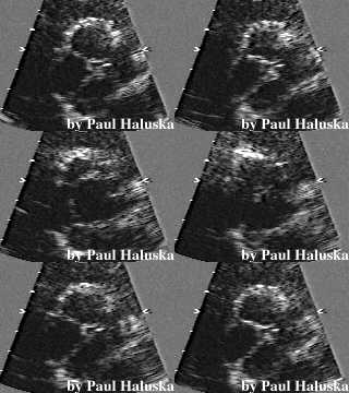
Quadricúspide

(Reported incidence of 0.008% to 0.033%. (European Heart Journal 1988;9:1269-1270)
.
Alguns achados são tão raros que passamos pela ecocardiografia sem ver um caso.
Semana passada acho que vi um, mas não tenho certeza.
Mas parecia com essas aqui.
.

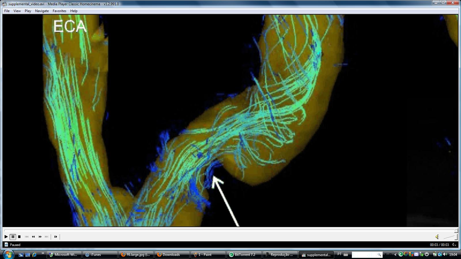
Tudo sobre os fluxos nas carótidas, e livre para baixar.

In Vivo Wall Shear Stress Distribution in the Carotid Artery

Effect of Bifurcation Geometry, Internal Carotid Artery Stenosis, and Recanalization Therapy

Conclusions— Flow-sensitive 4D MRI identified alterations in the segmental in vivo WSS distribution associated with atherosclerotic disease, surgical therapy, and individual bifurcation geometry and could be a valuable technique to assess the individual risk of flow-mediated atherosclerosis and carotid plaque progression. 

Artigo livre de estudos dos fluxos nas carótidas

E livre para baixar!

Não perca o filme, é demais.