Identification of Viable Myocardium in Acute Anterior Infarction Using Duration of Systolic Lengthening by Tissue Doppler Strain: A Preliminary Study
Results
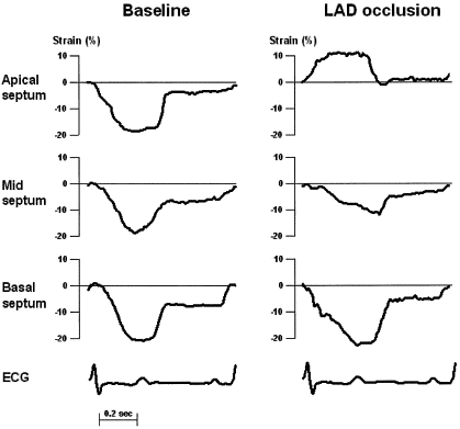
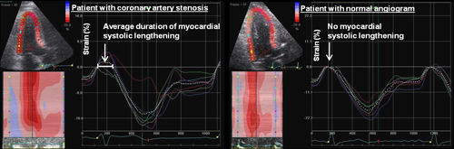
Peak systolic strain fell gradually (becoming less negative) from normal segments to segments with transmural infarction (P < .0001), and the duration of systolic lengthening increased (P < .0001). Myocardial scarring was closely correlated with peak systolic strain (R = 0.76, P < .00001) and the duration of systolic lengthening (R = 0.88, P < .00001). There was a significant correlation between the degree of scarring and time to percutaneous coronary intervention (R = 0.40, P = .045). In segments with systolic lengthening, the improvement in strain after remodeling was significantly higher (5.5 ± 5.1%) than in segments with duration of systolic lengthening > 67% of systole (2.2 ± 3.7%) (P 67.3% could identify nonviable myocardium (sensitivity, 90%; specificity, 94%).
Conclusions
In patients with acute myocardial infarctions in the anterior wall, strain measurements can identify myocardium with nontransmural scarring. The duration of systolic lengthening is a novel, easily implemented variable that may identify ischemic but viable myocardium. Myocardial infarctions in other left ventricular regions should be investigated in future studies.
.
.
A duração do Strain revela mais sobre a viabilidade do tecido do que se imaginava.
Será capaz de dar a viabilidade já na fase aguda?
.
É incrível como o Strain renovou a pesquisa em ecocardiografia e colocou o método de volta ao centro das atenções em cardiologia!
.