.
Loss of Adrenergic Augmentation of Diastolic Intra-LV Pressure Difference in Patients With Diastolic DysfunctionEvaluation by Color M-Mode Echocardiography
.
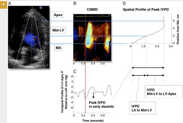
Methods We studied 166 consecutive patients undergoing dobutamine stress echocardiography who had no inducible ischemia and an EF ≥50%, of which 21 had normal diastolic function, 14 had impaired relaxation (grade 1), 80 had pseudonormal filling (grade 2), and 51 had restrictive filling (grade 3). Color M-mode Doppler (CMMD) images of mitral inflow were obtained at rest and during low (10 μg/kg/min) and peak (20 to 40 μg/kg/min) doses of dobutamine. The total IVPD from the LA to LV apex, LA to mid-LV, and mid-LV to the LV apex were calculated using the CMMD data to integrate the Euler equation.
Results Total IVPD was not different between groups at rest. With dobutamine, the total IVPD increased by 2.20 ± 1.95 mm Hg in normal subjects and by only 0.73 ± 1.33 mm Hg, 1.84 ± 1.63 mm Hg, and 1.08 ± 1.57 mm Hg in patients with grades 1, 2, and 3 DD, respectively. This difference was due to a failure in augmentation of IVPD from the mid-LV to the LV apex, indicating reduced apical ventricular suction with DD, whereas the IVPD from the LA to the mid-LV responded similarly to dobutamine in normal subjects and those with DD.
.
Conclusions In patients with preserved EF, DD is associated with a reduced adrenergic augmentation of the IVPD from the mid-LV to the LV apex, reflecting less apical suction.
.
Você chegou a pensar que o modo M Color não servia para mais nada?
.
Interessante trabalho publicado no JACC mostra variações do gradiente entre o átrio e ventrículo na diástole e a ação da Dobutamina sobre ele.
.
Parece que o ápice perde mesmo a sucção induzida por estímulo adrenérgico.
.
Este estudo ajudou a esclarecer uma dúvida:
Como a velocidade da onda E era aumentada na taquicardia fisiológica para acelerar o fluxo mitral passivo com o encurtamento da diástole?
.
Afinal, a taquicardia do exercício encurta o tempo de enchimento, uma condição que poderia piorar o débito.
.
Caso houve um aumento do gradiente, isso só poderia ser causado pela elevação das pressões atriais.
Que transmitidas aos capilares, poderia prejudicar a troca alveolar.
.
A resposta está na sucção do sangue do átrio pelo ventrículo esquerdo.
Sucção essa derivada da energia armazenada na sístole e liberada na diástole.
.
Sucção também conhecida como onda e´ !!!!
.
O estresse sistólico aumentado na taquicardia fisiológica provoca uma sucção elevada.
.
Você chegou a pensar que o modo M Color não servia para mais nada?
.
Interessante trabalho publicado no JACC mostra variações do gradiente entre o átrio e ventrículo na diástole e a ação da Dobutamina sobre ele.
.
Parece que o ápice perde mesmo a sucção induzida por estímulo adrenérgico.
.
Este estudo ajudou a esclarecer uma dúvida:
Como a velocidade da onda E era aumentada na taquicardia fisiológica para acelerar o fluxo mitral passivo com o encurtamento da diástole?
.
Afinal, a taquicardia do exercício encurta o tempo de enchimento, uma condição que poderia piorar o débito.
.
Caso houve um aumento do gradiente, isso só poderia ser causado pela elevação das pressões atriais.
Que transmitidas aos capilares, poderia prejudicar a troca alveolar.
.
A resposta está na sucção do sangue do átrio pelo ventrículo esquerdo.
Sucção essa derivada da energia armazenada na sístole e liberada na diástole.
.
Sucção também conhecida como onda e´ !!!!
.
O estresse sistólico aumentado na taquicardia fisiológica provoca uma sucção elevada.
.