
_uacct = “UA-1399567-1”;
urchinTracker();
Mês: junho 2013
Transdutor: Cuidados com seu patrimônio

.
Cabos de transdutores NUNCA devem tocar o chão, serem pisados ou enrolarem nas rodas.
Fratura de cabo é prejuízo grande e certo.
Ensine suas funcionárias!
.
.
Papel áspero NUNCA deve ser usado na borda de pele do transdutor. Raspam a membrana e favorecem as rachaduras.
.
O e,i e q do Vivid GE

Os leitores do blog frequentemente perguntam de portáteis.
Como a GE domina amplamente esse mercado, as dúvidas são sempre:
Vivid “e” ou Vivid “i”?
Vivid “i” ou Vivid “q”???
Quando a questão é Vivid “e” ou Vivid “i”, é mais fácil responder.
Qual é a expectativa de movimento? Fará exames a beira do leito em UTI?
Com um movimento acima de 120 ecos por mês e casos hospitalares, a melhor escolha é o “i”.
Para casos de convênios ou SUS ambulatorial, o “e” dá e sobra.
Já orientar sobre o Vivid “i” ou Vivid “q” é mais complexo.
Depende muito do futuro do Strain.
Acreditando na tecnologia, tanto quanto o Castilho e Ronaldo, a compra adequada é o “q”.
Em curto prazo, fazendo rotina diagnóstica com pagamento por convênios, o “i” é mais custo-eficiente.
Nós aqui da EchoTalk? Compraríamos o “q”.
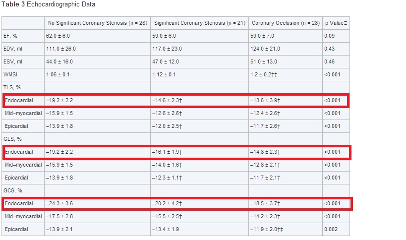
A isquemia que pode ser quantificada ao Eco-Strain está no endocárdio e não depende de bordas.
.
Caso você não tenha se empolgando com o estudo do post anterior, AQUI, reveja pelo menos a tabela acima.
Foi um estudo em hospital, com pacientes com síndrome coronária sem supra, casos que ocorrem aos milhares nesse minuto em todo o Brasil.
.
Para quem desligou o raciocínio clínico e pede cateterismo para todos, o impacto será zero.
.
Em serviços que evitam exames desnecessários, essa pode ser a saída.
Um exame de ecocardiograma de 20 minutos com até a possibilidade de avaliação dos resultados à distância…
.
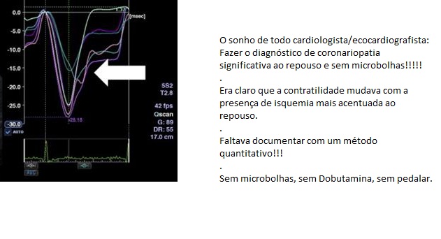
Notem como a porção endocárdica, que já é parcialmente isquêmica, fica pior ainda e contrai muito pouco.
.
Poderoso Thor: Diagnóstico de coronariopatia ao repouso por Strain. Pesquisa do ano de 2013 faz o funeral do contraste.
Layer-Specific Quantification of Myocardial Deformation by Strain Echocardiography May Reveal Significant CAD in Patients With Non–ST-Segment Elevation Acute Coronary Syndrome
.
Prof. Thor Edvardsen, Department of Cardiology, Oslo University Hospital, Rikshospitalet, N-0027 Oslo, Norway
.
.
Seventy-seven patients referred to coronary angiography due to suspected non–ST-segment elevation-acute coronary syndromes (NSTE-ACS) were prospectively included. Coronary occlusion was found in 28, significant stenosis in 21, and no stenosis in 28 patients. Echocardiography was performed 1 to 2 h before angiography. Layer-specific longitudinal and circumferential strains were assessed from endocardium, mid-myocardium, and epicardium by 2-dimensional (2D) speckle-tracking echocardiography (STE). Territorial longitudinal strain (TLS) was calculated based on the perfusion territories of the 3 major coronary arteries in a 16-segment LV model, whereas global circumferential strain (GCS) was averaged from 6 circumferential LV segments in all 3 layers.
.
Results Patients with significant CAD had worse function in all 3 myocardial layers assessed by TLS and GCS compared with patients without significant CAD. Endocardial TLS (mean –14.0 ± 3.3% vs. –19.2 ± 2.2%; p < 0.001) and GCS (mean –19.3 ± 4.0% vs. –24.3 ± 3.4%; p < 0.001) were most affected. The absolute differences between endocardial and epicardial TLS and GCS were lower in patients with significant CAD (Δ2.4 ± 3.6% and Δ6.7 ± 3.8%, respectively) than in those without significant CAD (Δ5.3 ± 2.1% and Δ10.4 ± 3.0%; p < 0.001). This reflects a pronounced decrease in endocardial function in patients with significant CAD
.
.
2D-STE
Grayscale images were analyzed. Myocardial function by strain was evaluated on a frame-by-frame basis by automatic tracking of acoustic markers (speckles) throughout the cardiac cycle. The endocardial borders were traced in the end-systolic frame of the 2D images from the 3 apical views for analyses of longitudinal endocardial, mid-myocardial, and epicardial strains. Analyses of layer-specific circumferential strains were obtained from the parasternal short-axis view. Peak negative systolic longitudinal and circumferential strains from 3 layers were assessed using off-line software (Toshiba Medical Systems Corporation, Tokyo, Japan) in 16 longitudinal and 6 circumferential LV segments. All segmental values were averaged to global longitudinal strain (GLS) and global circumferential strain (GCS) for each myocardial layer (Figure 1). Segments that failed to track properly were manually adjusted by the operator. Any segments that subsequently failed to track were excluded.
.
Toshiba?!?!?!?!?!? Quem diria…
.
.
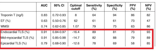
Então veio de um pesquisador da Noruega, chamado Thor, usando um Toshiba(?), sem contraste e com uma sensibilidade igual a da tomografia 64(+) ?????????!!!!!!!!!!
.
Absolutamente surpreendente.
.