3D SC 2000: A máquina vence o homem

Ontem fizemos um exame exemplar.
Feito pela residente Mari, o exame parecia normal mas o 3D do ventrículo apresentava fração de ejeção rebaixada.
Fui rever o exame e também achei que fosse normal, mesmo repetindo o 3D do VE e a fração de ejeção ficando em 45%!
.
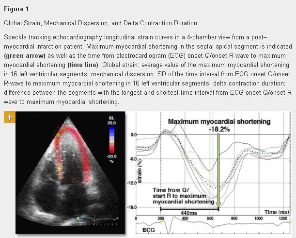
Aqui no blog foram publicados estudos de 3D e Strain que afirmavam categoricamente:
As novas tecnologias ajudam o médico inexperiente, não ajudam o experiente.
.
Após 18 anos de ecocardiografia, posso ser considerado experiente.
.
Voltamos à máquina e refizemos tudo. A análise segmentar do volume mostrava as porções média e basal lateral como hipocinéticas.
.
O volume 3D mostrava mesmo a região sem a contratilidade esperada…
Estávamos errados e o 3D SC 2000 nos corrigiu!!!!
.
Veja outro caso aqui

.

.
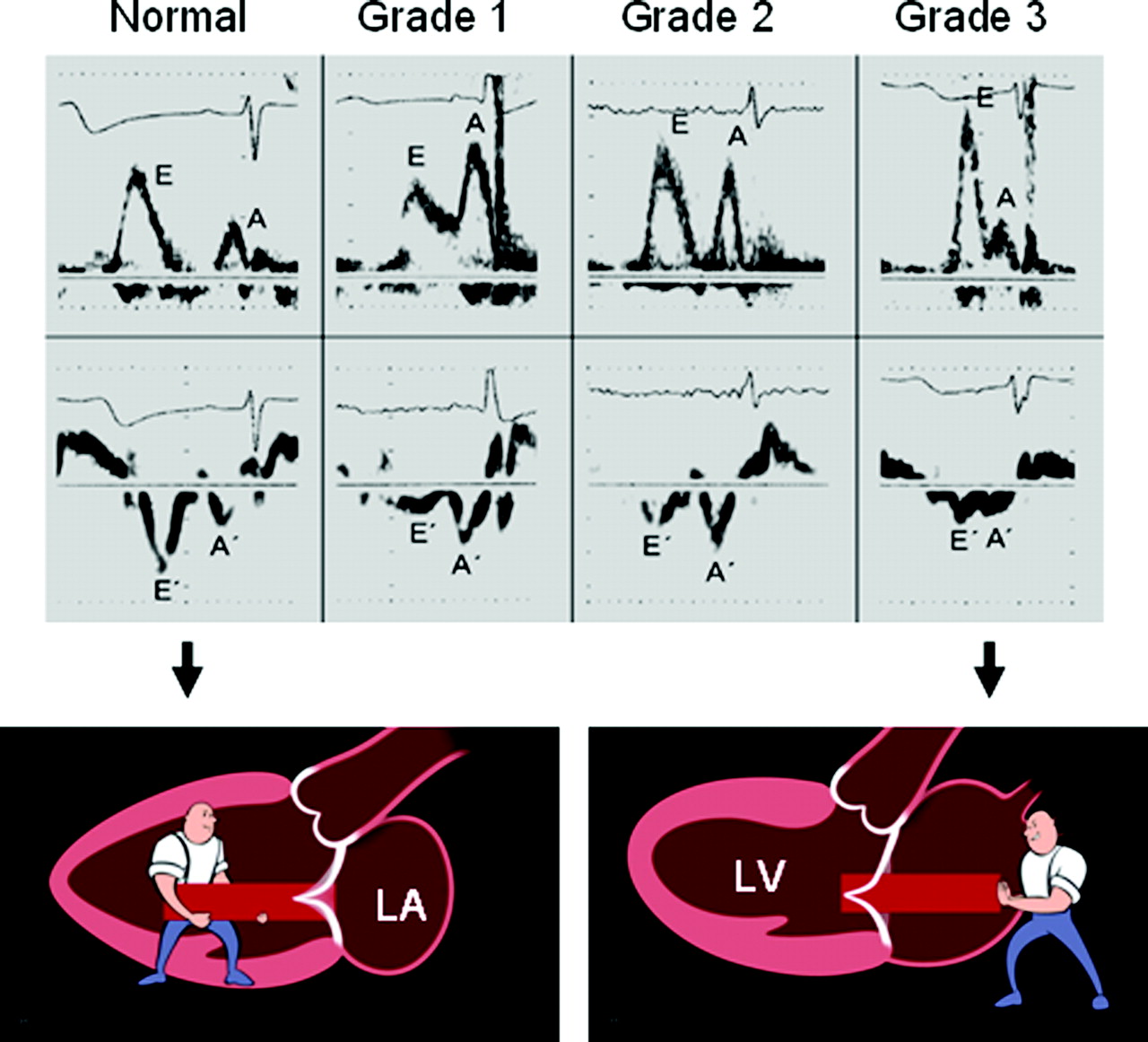
Diastologia é difícil? Não para o J. Oh.


.
Aqui artigo de mestre completo e irresistível!!!!
.
Ladies and gentleman, I tried to summarize for you today in my lecture the current clinical use of echocardiography in patients with heart failure, not only systolic but also diastolic heart failure. There are many exciting new technologies and applications of echocardiography such as 3-dimensional echocardiography, strain imaging, and myocardial perfusion imaging. I am certain that these new applications will strengthen and improve the role of echocardiography in general, but more so in patients with symptoms of heart failure. Undoubtedly, echocardiography is the single most useful test in patients with symptoms of heart failure. It is essential in the diagnosis and identification of underlying etiology of heart failure. However, we should move beyond the concept of echocardiography as only a diagnostic tool in patients with heart failure, hence, use this versatile and easily available technology to gain pathophysiologic insights of various forms of heart failure, to help identify or establish an optimal therapy for the patients with heart failure, to monitor treatment response, to prognosticate, and to be an important tool in clinical heart failure trials.

Color 3D: Novo padrão ouro de refluxo

Aqui artigo
.

.
Aqui aula ótima
.
O ColorDoppler sempre foi um perigo para as avaliações de refluxo valvar. O efeito bola de bilhar das hemáceas, empurrando as outras e fazendo imagens enormes de mosaico ao Color já fizeram muitas avaliações naufragarem. Isso não ocorre ao 3D color no local de refluxo e a área pode ser medida com precisão