.
.

.
.
.
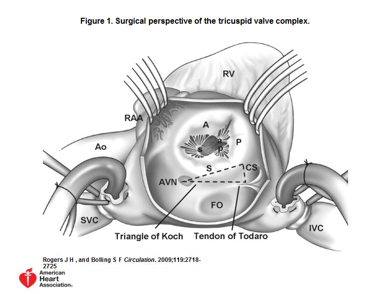
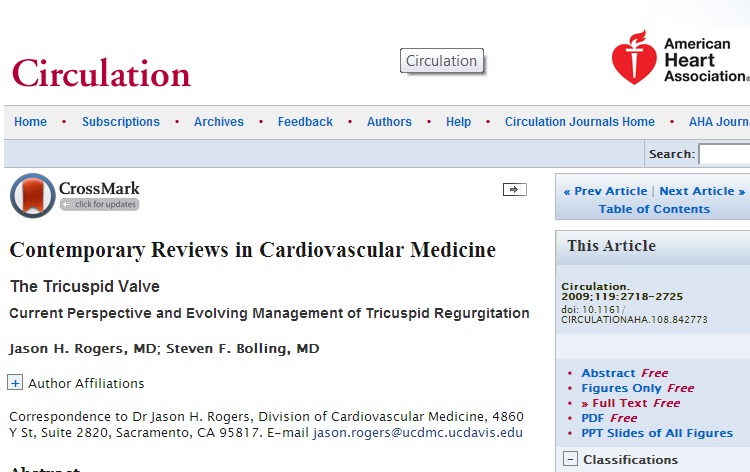
Cardiovascular specialists have entered an era of renewed interest and enthusiasm surrounding the diagnosis and treatment of valvular heart disease, driven in part by emerging percutaneous therapies for the treatment of aortic, pulmonic, and mitral valve disease. Despite this wave of investigation, little or no attention has been given to the treatment of tricuspid valve disease
.
As válvulas estão voltando.
Sem a febre reumática, as valvopatias perderam espaço nos estudos médicos.
Agora, as novas técnicas de implante, o envelhecimento da população e a alta prevalência de coronariopatas crônicos nos levam de volta ao centro da questão:
Ecocardiografia de lesões valvares.
E não esqueçam da Tricúspide!!
.
.
.
 .
.
.
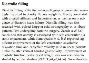
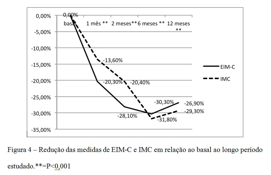
Nosso grupo estuda os efeitos da cirurgia bariátrica no coração e carótidas.
Podemos adiantar que os benefícios são bem mais precoces que a perda de peso efetiva.
Vejam no congresso a apresentação abaixo
.
.
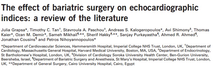
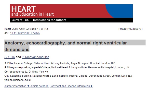
Quem melhor que o Nihoyannopoulos para falar sobre o ventrículo esquecido, o direito?
Artigo de revisão muito útil para os ecocardiografistas
.
Devemos escapar dos laudos genéricos sobre o ventrículo direito e passarmos a avaliações precisas do volume e função.
.
 .
.
.
.
O blog já alertou sobre o uso crescente do ecocardiograma pela equipe de quimioterapia.
Não basta realizar um exame básico.
Veja no texto.
.
 .
.
.
.
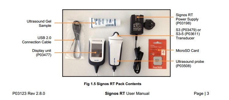
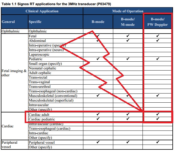
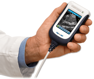
Já vi muitos ecocardiografistas criticarem os ultra portáteis por não terem Doppler pulsado.
Inclusive meu guru Morcerf!
.
Agora já temos aparelhos com Doppler.
Que boa notícia!!!
.
.
.
.
.
.
Considering the observations that RWMAs are an early sign of myocardial ischemia and that the absence of RWMAs effectively rules out ischemia, a group of investigators prospectively studied the utility of echocardiography in the triage of patients with chest pain [5]. Among 180 consecutive patients presenting to the emergency department at an academic hospital who met inclusion criteria, 94 percent had adequate echocardiograms. Sixty of 169 (36 percent) had no regional or global dysfunction and 22 (13 percent) had global hypokinesis without RWMAs. The following results were noted:
●Of the 88 patients without RWMAs, only 2 (2 percent) subsequently “ruled in” for a non-Q wave myocardial infarction by cardiac enzymes, without clinical sequelae (figure 2).
●Of the 87 patients with RWMAs, 27 (31 percent) had an acute myocardial infarction. The initial ECG was diagnostic (ST elevation with or without Q waves) in nine of these patients, and eight had an electrocardiogram that was not interpretable for an acute MI (due to LBBB, pacing, or left ventricular hypertrophy with strain). Thirteen patients subsequently developed pathologic Q waves.
Thus, one strategy for screening chest pain patients for RWMAs with echocardiography would be as follows.
●If an adequate echocardiogram can be obtained and there are no RWMAs, it is relative safe to discharge the patient from the emergency department.
●Patients with RWMAs should be admitted for further observation and treatment. Those patients with a suspicious chest pain syndrome in whom an adequate echocardiogram cannot be obtained would also warrant observation.
.
 .
.
.
Muito bom acertar em 96% dos casos com o ecocardiograma no pronto socorro.
.
 .
.
.
Link para entrevista do administrador
.
.
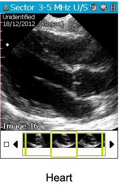
Emergency and Intensive Care physicians can assess their patients in the department saving time and improving patient management rather than waiting to transport the patient to and from the ultrasound department.
.
.
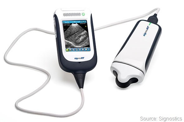
The screen is a touch screen and practitioners can either navigate the tool bar with the stylus or by using the scroll wheel. B-mode, M-mode and Pulse Wave Doppler are included and multiple presets are available to optimise the image easily.
.
.
.
So, deformation stress echo appears feasible, is able to identify pathology with as good if not better sensitivity than traditional wall motion analysis, and does appear to offer additional prognostic information to that gained by wall motion assessment alone. However, stress echo by any method has a steep learning curve and significant experience is needed in order to maximize its sensitivity. Unfortunately, these new tools at present may not significantly reduce this as the advanced echo techniques themselves demand a degree of expertise. There is also the time factor to overcome, but some of the newer more automated techniques may help in both these regards. In addition, appropriate cut-off values for normality vs. different types of abnormality (ischaemia, viability, etc.) need to be defined from a detailed meta-analysis of the literature, as currently there are no clear definitions and each study has used their own values according to varying endpoints.