Enxergar a coronária é isso!

In-vivo optical coherence tomography image of a ruptured coronary plaque. Ruptured plaque (RP) is seen in this patient with severe disruption of plaque fibrous cap at the 6 o’clock position. Adherent thrombus (Thr) is also observed at 1 o’clock position. Guidewire artifact is represented by GW.

Association of statin therapy with reduced coronary plaque rupture: an optical coherence tomography study
Volume 19(4), June 2008, pp 237-242

Sim, o ideal seria obter uma imagem dessa de forma não invasiva.
Mas enquanto não é possível, e a Tomo 65 está longe disso, veja que imagem elegante da placa, com rutura e um trombinho!

Espessamento da carótida: Dislipidemia na infância, Aterosclerose na maturidade


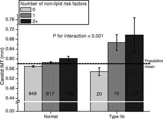
Associations of Dyslipidemias From Childhood to Adulthood With Carotid Intima-Media Thickness, Elasticity, and Brachial Flow-Mediated Dilatation in Adulthood: The Cardiovascular Risk in Young Finns Study

Volume 28(5), May 2008, pp 1012-1017
Background—: Dyslipidemias are the major cause for atherosclerosis. They may act synergistically with nonlipid risk factors to increase atherogenesis. In the present study, we examined the effects of dyslipidemias from childhood to adulthood and their interaction with nonlipid risk factors on markers of subclinical atherosclerosis.

Methods and Results—: Study subjects were participants of the longitudinal Cardiovascular Risk in Young Finns Study started in 1980 (n=2265, age 3 to 18 years). To phenotype type IIa, IIb, and IV dyslipidemias and hypoHDL-cholesterolemia, we calculated age and sex-specific z scores for lipid values for each subject in 1980, 1983, 1986, and 2001. Subjects with mean z score over 90th percentile for LDL-cholesterol or triglycerides were considered having type IIa or IV dyslipidemia. Subjects with mean z score over 90th percentile for LDL-cholesterol and triglycerides had type IIb dyslipidemia, and those with mean z score below 10th percentile for HDL-cholesterol had hypoHDL-cholesterolemia. Compared to controls, subjects with type IIb dyslipidemia had increased carotid IMT (P<0.01). This difference remained significant when adjusted with other risk factors (P<0.05). Carotid IMT also increased significantly more with increasing number of nonlipid risk factors (P<0.001) or presence of the metabolic syndrome (P<0.05) in subjects with type IIb than in controls. Subjects with type IIb or type IV dyslipidemia had decreased carotid elasticity (P0.3) when adjusted with blood pressure.

Conclusions—: Our findings suggest that type IIb dyslipidemia has deleterious effects on vasculature already since childhood. Subjects with type IIb dyslipidemia are more vulnerable to the effects of cardiovascular risk factors and metabolic syndrome.

Aterosclerose:Regressão , progressão ou manutenção


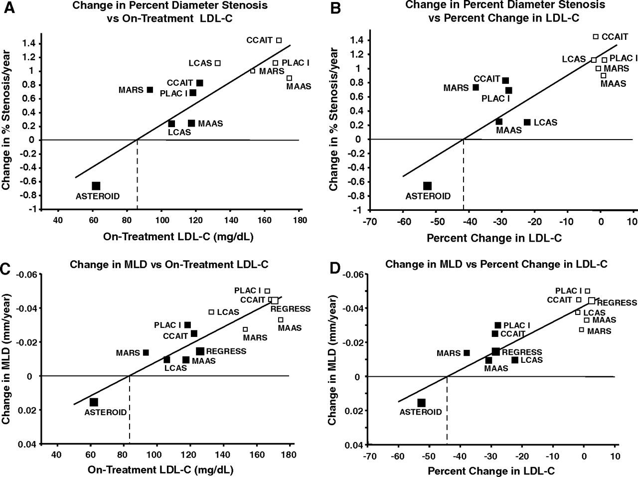
Effect of Rosuvastatin Therapy on Coronary Artery Stenoses Assessed by Quantitative Coronary Angiography
A Study to Evaluate the Effect of Rosuvastatin on Intravascular Ultrasound-Derived Coronary Atheroma Burden
Circulation. 2008;117:2458-2466

Mais um esforço para metas de colesterol abaixo de 70mg/dl!
Detalhes:
O estudo envolvia apenas pacientes com placas maiores que 20% e menores Que 50% de estenose, isto é, placas moles!
Usou o parâmetro de mínimo lumem para definir melhora, mas se tem algo que muda em uma artéria é seu lumem!
Fazendo estudos de reatividade vascular vejo variações impressionantes em segundos.
Veja o filme da carótida acima, não é só colesterol o problema!

Ressincronização: nem modo-M, nem Simpson.

Quando testamos através da fração de ejeção a eficácia da ressincronização ventricular, cometemos um engano.
Mesmo o Simpson, mas ainda mais o modo-M, representam um instante fotográfico da sístole e diástole.
Com a assincronia do BRE, esse momento não registrará o volume ejetado de fato.
Com a ressincronização, a foto registrará o momento de menor volume e o de maior volume ventricular, dando a incompleta interpretação de melhora.
Apenas o cálculo do volume ejetado do VE deve ser usado, com base no VTI da via de saída e sua área. Pois ele independe do exato instante da contração, representa a ejeção claramente.
Com o volume ejetado, é só fazer o cálculo do volume diastólico final ao 4 e 2 câmaras para calcular a fração de ejeção e sua “possível” melhora.

Ressincronização: Verdades e mentiras


Relationship Between Intraventricular Cardiac Asynchrony and Degree of Systolic Dysfunction

Journal of the American Society of Echocardiography

Volume 21(3), March 2008, p 214–218

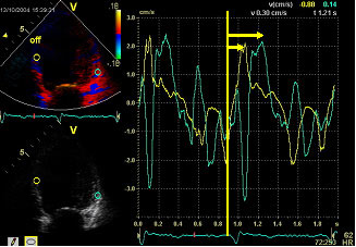
Objectives: Cardiac asynchrony is an area of study becoming more relevant in the evaluation and management of heart failure. Our aim was to determine the prevalence of cardiac asynchrony by Doppler echocardiography and to evaluate its relationship with the degree of left ventricular (LV) systolic dysfunction.

METHODS: A total of 316 consecutive patients with LV ejection fraction less than 40% were enrolled. We divided them into 3 groups according to the degree of LV dysfunction: 31% to 40%, 21% to 30%, and less than 20%. Intraventricular asynchrony was evaluated using two methods: (1) measurement of the septal to posterior wall-motion delay (cut-off point 130 milliseconds); and (2) measurement of the difference between time from Q wave to LV ejection end, and the time from Q wave to the end of the systolic wave of the most delayed basal segment by Doppler tissue imaging (ejection- Doppler tissue imaging time; cut-off point 50 milliseconds).

RESULTS: Mean age was 62.14 ± 13.5 years (75.7% men). No differences were found among clinical electrical and echocardiographic variables among the groups. Furthermore, no relationship was found between the existence of intraventricular cardiac asynchrony and the degree of LV systolic dysfunction. These were similar in patients with ischemic dilated cardiomyopathy and nonischemic dilated cardiomyopathy.

The degree of LV systolic dysfunction and its origin are not related to the presence of cardiac asynchrony. A specific echocardiographic Doppler study must always be performed to assess the existence of cardiac asynchrony in those who are candidates to resynchronization therapy.<span

Atração (?!?) do DEPECO 2008


Sessão “Você é o Cirurgião”
Descrição

Demonstração prática com dissecção em coração de porco, orientada por cirurgiões cardíacos. Esta atividade visa a compreensão da anatomia ecocardiografica e das principais técnicas cirúrgicas. Será dada ênfase nas valvopatias e circulação venosa coronariana (colocação de eletrodo de marcapasso para ressincronização venticular).

30 Vagas cada Sessão

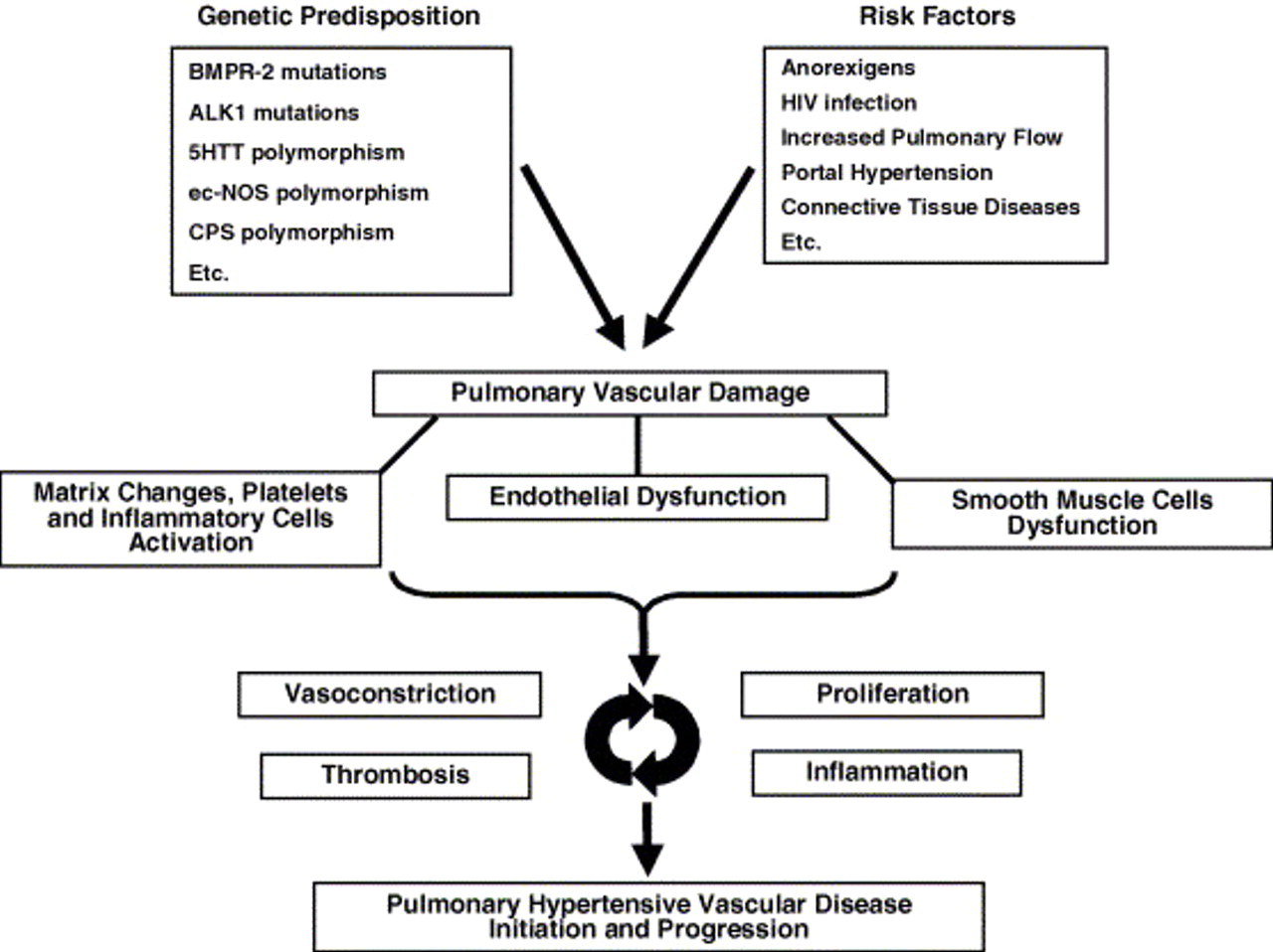
HIPERTENSÃO PULMONAR


Guidelines on diagnosis and treatment of pulmonary arterial hypertension
The Task Force on Diagnosis and Treatment of Pulmonary Arterial Hypertension of the European Society of Cardiology
European Heart Journal 2004 25(24):2243-2278

According to these data mild PH can be defined as a PASP of approximately 36–50 mmHg

Os guidelines clínicos dividem a hipertensão pulmonar em leve ou significativa, sem classificá-la como moderada.
Para medida da pressão estimada ao ecocardiograma,
36 a 50mmhg é leve, acima disso é significativa, importante ou acentuada.

IMT em pacientes com Lesão medular: Atherosclerosis 2008


Matos-Souza JR, et al., Carotid intima-media thickness is increased in patients with spinal cord injury independent of traditional cardiovascular risk factors, Atherosclerosis (2008)
In summary, the present report demonstrated that SCI
males with markers of low cardiovascular risk had higher carotid IMT than able-bodied controls, independent of traditional risk factors clustering. Thus, alternative mechanisms such as increased inflammation and endothelial activation
seen in injured patients might potentially play a role
in this process
.