
Differential expression of cytokines, chemokines and chemokine receptors in patients with coronary artery disease.
Int J Cardiol. 2008 Jul 8.
Oliveira RT, Mamoni RL, Souza JR, Fernandes JL, Rios FJ, Gidlund M, Coelho OR, Blotta MH.
Department of Clinical Pathology, Faculty of Medical Sciences, State University of Campinas (UNICAMP), Campinas, SP, Brazil.
.
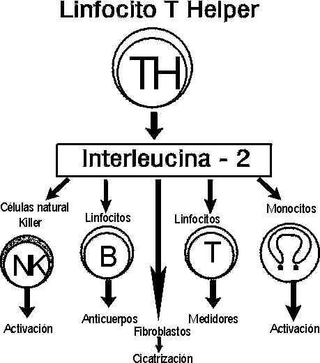
Monocytes/macrophages and lymphocytes have a key role in the pathogenesis of atherosclerosis through the production of inflammatory and anti-inflammatory cytokines. We evaluated mRNA expression and protein production of CCL2, CXCL8, CXCL9, CXCL10, IFN-gamma and IL-10 in vitro as well as the expression of the CCR2 and CXCR3 receptors in peripheral blood mononuclear cells (PBMCs) of patients with coronary artery disease (CAD) and healthy controls in the presence or absence of oxidized LDL (oxLDL). Patients with CAD showed higher constitutive expression of CCL2, CXCL8, CXCL9, CXCL10 and IFN-gamma mRNA and, after stimulation with oxLDL, higher expression of CCL2 and CXCL8 mRNA than the control group. We also detected higher levels of CCL2 and CXCL8 in supernatants of oxLDL-stimulated PBMCs from CAD patients than in corresponding supernatants from controls. Patients with CAD had a higher percentage of constitutive CCR2(+) and CXCR3(+) cells after stimulation with oxLDL. Among CAD patients, the main differences between the stable (SA) and unstable angina (UA) groups were lower IL-10 mRNA production in the latter group. Altogether, our data suggest that PBMCs from CAD patients are able to produce higher concentrations of chemokines and cytokines involved in the regulation of monocyte and lymphocyte migration and retention in atherosclerotic lesions.
.
Continuamos nossa pesquisa da fisiopatologia da Aterosclerose. Além de marcadores séricos de inflamação, como interleucina-6, demonstramos a ativação dos linfócitos.
Não há mais duvida, o alarme inflamatório se liga intimamente aos eventos agudos da síndrome coronária.
Dia: 21 de janeiro de 2009
Você está pronto para o 3D de rotina?
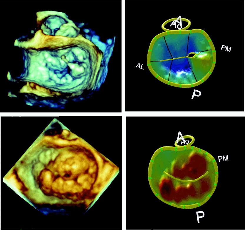
Real-Time 3-Dimensional Echocardiography: An Integral Component of the Routine Echocardiographic Examination in Adult Patients?
Circulation Volume 119(2), 20 January 2009, pp 314-329
.
“Currently, many laboratories perform a complete 2DE study, followed by a focused 3D examination, in patients with specific pathologies in which RT3DE imaging could potentially provide additional diagnostic information. It can be anticipated that a full-volume acquisition of the left ventricle will be performed in every patient to obtain LV volumes and EF. The 3D images should be stored in a digital archiving system with the 2D study to allow integrated interpretation of all images and incorporation of 3D findings into the report.
Future advances in transducer and computer technology will result in several important improvements that will further enhance the clinical application of RT3DE imaging. One highly desirable improvement is the ability to acquire wider-angle pyramid of data with and without color flow in a single cardiac cycle. This will shorten data acquisition and eliminate stitching artifacts. Furthermore, future improvements in both spatial resolution and temporal resolution of the transthoracic RT3DE imaging, which are still below those of 2DE, will broaden the spectrum of patients who can be imaged with this modality. Further miniaturization of the 3D MTEE technology will allow 3D TEE imaging in pediatric patients and the development of real-time 3D imaging intracardiac catheters. Future software developments will allow new types of sophisticated quantitative analysis of the cardiovascular anatomy and function, including the fusion of RT3DE data with other 3D imaging modalities such as magnetic resonance and computed tomography.”
.
A pergunta não é se o 3D já está pronto, e sim se nós estamos prontos para o 3D.
Insisto que usar o 3D após o exame em 2D é um erro, e prejudica a ampla diusão do método.
É para usar o 3D do início ao fim.
Sempre ele, o volume atrial.
Left atrial remodelling in patients with myocardial infarction complicated by heart failure, left ventricular dysfunction, or both: the VALIANT Echo Study
European heart journal Volume 30(1), January 2009, pp 56-65
.
Aims: To assess the relationship between left atrial (LA) size and outcome after high-risk myocardial infarction (MI) and to study dynamic changes in LA size during long-term follow-up.
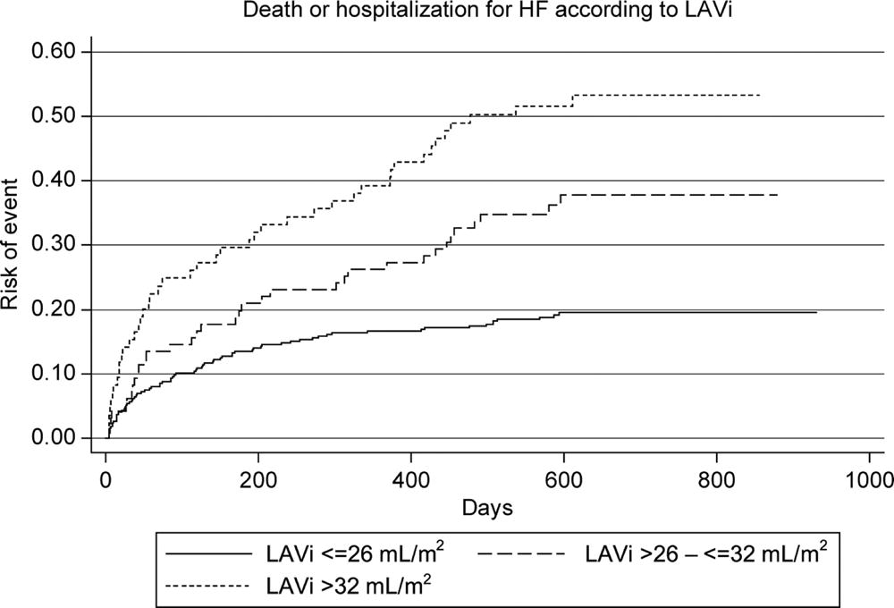
Methods and results: The VALIANT Echocardiography study prospectively enrolled 610 patients with left ventricular (LV) dysfunction, heart failure (HF), or both following MI. We assessed LA volume indexed to body surface area (LAVi) at baseline, 1 month, and 20 months after MI. Baseline LAVi was an independent predictor of all-cause death or HF hospitalization (P=0.004). In patients who survived to 20 months, LAVi increased a mean of 3.00 ± 7.08 mL/m2 from baseline. Hypertension, lower estimated glomerular filtration rate, and LV mass were the only baseline independent predictors of LA remodelling. Changes in LA size were related to worsening in MR and increasing in LV volumes. LA enlargement during the first month was significantly greater in patients who subsequently died or were hospitalized for HF than in patients without events.
Conclusion: Baseline LA size is an independent predictor of death or HF hospitalization following high-risk MI. Moreover, LA remodelling during the first month after infarction is associated with adverse outcome.
.
O volume atrial é o melhor marcador de disfunção diastólica e um ótimo índice de prognóstico.
Nesse artigo, fica evidente que é um marcador precoce de má evolução.