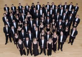
EchoTalk é um grupo.

.
O retorno ao blog ficou mais saboroso agora que dividimos as postagens, inicialmente com o Dr. Guilherme e mais para a frente com os outros membros da EchoTalk.
.
A força de um grupo unido, supera qualquer estrela nacional da ecocardiografia.
Abraço a todos e vamos deixar 2011 ser o que promete.
.
Beto

FREUD EXPLICA?

“CONTRA OS ATAQUES NÓS PODEMOS NOS DEFENDER!

CONTRA OS ELOGIOS NÃO HÁ NADA QUE POSSAMOS FAZER…”
-Sigmund Freud-
FIQUEI MUITO FELIZ AO SABER QUE HÁ ESCOLAS DE ECOCARDIOGRAFIA E DOPPLER DA CAPITAL OFERECENDO CURSOS “HANDS ON” (ISSO MESMO, “HANDS ON”!!!).
SENDO UMA TENDÊNCIA INICIADA PELO PROFESSOR JOSÉ ROBERTO AQUI NA ECHOTALK, É BOM SABER QUE NOSSA CRUZADA CONTRA OS CURSOS EM POWER POINT ESTÁ SURTINDO EFEITO NA CONCORRÊNCIA!
BOM PARA OS ALUNOS…

DEU NO CIRCULATION!


Multimodality Cardiac Imaging in the Evaluation of Mitral Annular Caseous Calcification

Gulati, Ankur MA, MRCP; Chan, Cheuk MRCP; Duncan, Alison MRCP, PhD; Raza, Sadaf BSc; Kilner, Philip J. MD, PhD; Pepper, John MA, FRCS

Author Information

From The Royal Brompton Hospital, South Kensington, United Kingdom, SW3 6NP.

The online-only Data Supplement is available with this article athttp://circ.ahajournals.org/cgi/content/full/123/1/e1/DC1.

Correspondence to Dr Ankur Gulati, Cardiac Magnetic Resonance Unit, The Royal Brompton Hospital, Sydney St, South Kensington, United Kingdom, SW3 6NP. E-mail a.gulati@rbht.nhs.uk

A 77-year-old man with severe aortic stenosis was referred for consideration of aortic valve replacement. He admitted to mild exertional dyspnea and was systemically well. His past medical history was remarkable for hypertension, hypercholesterolemia, and a previous transient ischemic attack. A transthoracic echocardiogram confirmed severe calcific aortic stenosis (peak gradient 95mm Hg, estimated orifice area 0.5 cm2) with normal left ventricular ejection fraction (70%). In addition, a well-circumscribed, echodense mass (25 mm in diameter) was observed in the posterolateral basal wall attached to the posterior mitral valve leaflet (Figures 1A and 1B). Although highly echogenic, the mass also contained central echolucent areas. The mitral valve was competent, with no functional stenosis. There was no pericardial effusion. Three-dimensional transesophageal echocardiography confirmed an encapsulated, heterogenous mass arising from the posterior mitral valve annulus, adherent to the posterior mitral valve leaflet P2 scallop, in close proximity to the left atrioventricular groove and circumflex artery (Figures 1C and 1D; online-only Data Supplement, Movie 1). The mass did not appear to be pedunculated or vascular in nature.

The patient was referred for cardiac magnetic resonance imaging to further characterize the cardiac mass. Cine imaging with a balanced steady state free-precession sequence showed an immobile, low-signal mass with a smooth contour located at the posterior mitral valve annulus (Figure 2A; online-only Data Supplement, Movie 2). There was no restriction of the posterior mitral valve leaflet motion or associated mitral regurgitation. On both multicontrast T1-weighted turbo spin-echo (Figure 2B) and short-tau inversion recovery T2-weighted images (Figure 2C), the mass was hypointense. After intravenous gadolinium contrast administration, first-pass perfusion images revealed no enhancement of the mass, in keeping with an avascular structure. On the early gadolinium-enhancement images, the mass remained hypointense (Figure 2D); however, the corresponding late gadolinium-enhancement images acquired 15 to 20 minutes later demonstrated a prominent rim of circumferential enhancement that surrounded a dark central core (Figures 2E and 2F). The characteristic low-signal intensity of the mass seen with multispectral cardiac magnetic resonance analysis and the strong peripheral late enhancement were consistent with a central area of caseous tissue surrounded by a fibrotic cap.

The anatomic location, heterogenous echocardiographic appearances, and cardiac magnetic resonance signal characteristics of the mass were therefore thought to support a diagnosis of mitral annular caseous calcification. This diagnosis was confirmed by cardiac computed tomography, which revealed that the mass consisted of an isodense central area with a hyperdense rim of peripheral calcification (Figure 3). Of note, the aortic valve leaflets were also heavily calcified. The patient was recommended to undergo aortic valve replacement but subsequently declined surgery.

Caseous calcification of the mitral valve is a rare form of mitral annular calcification that typically affects the posterior annulus. The condition, also referred to as liquefaction necrosis, usually runs a benign course, although associated valvular dysfunction and cerebral embolization of necrotic material have both been reported. Appreciation of this unusual condition is important because the echocardiographic appearances are nonspecific and commonly misinterpreted. Differential diagnoses include cardiac tumors and myocardial abscess, with the result that accurate assessment of this pathology frequently requires complementary imaging techniques. To the best of our knowledge, this report is the first to comprehensively illustrate the characteristic appearances in a single case by use of the full spectrum of noninvasive cardiac imaging modalities (transthoracic and transesophageal echocardiography, cardiac magnetic resonance imaging, and cardiac computed tomography).

XXXXXXXXXXXXXXXXXXXXXXXXXXXXXXXX

TUDO BEM, BACANA! FIZERAM UM MONTE DE IMAGENS E TUDO…

MAS NO MUNDO REAL, SÓ O ECOCARDIO JÁ RESOLVERIA A VIDA DO PACIENTE, NÃO?!

ATEROSCLEROSE E CARÓTIDAS – MAIS EVIDÊNCIAS!

Association between Carotid Intima-Media Thickness and Aortic Arch Plaques

Tessitore, Elena MDa; Rundek, Tatjana MDc; Jin, Zhezhen PhDb; Homma, Shunichi MDa; Sacco, Ralph L. MDc,d; Di Tullio, Marco R. MDa


Background: Large aortic arch plaques are associated with ischemic stroke. Carotid intima-media thickness (CIMT) is a marker of subclinical atherosclerosis and a strong predictor of cardiovascular disease and stroke. The association between CIMT and aortic arch plaques has been studied in patients with strokes, but not in the general population. The aim of this study was to investigate this association in an elderly asymptomatic cohort and to assess the possibility of using CIMT to predict the presence or absence of large aortic arch plaques.

Methods: Stroke-free control subjects from the Aortic Plaque and Risk of Ischemic Stroke (APRIS) study underwent transesophageal echocardiography and high-resolution B-mode ultrasound of the carotid arteries. CIMT was measured at the common carotid artery, bifurcation, and internal carotid artery. The association between CIMT and aortic arch plaques was analyzed using multivariate regression models. The positive and negative predictive values of CIMT for large (>=4 mm) aortic arch plaques were calculated.

Results: Among 138 subjects, large aortic arch plaques were present in 35 (25.4%). Only CIMT at the bifurcation was associated with large aortic arch plaques after adjustment for atherosclerotic risk factors (P = .007). The positive and negative predictive values of CIMT for aortic arch plaque >= 4 mm at the bifurcation above the 75th percentile (>=0.95 mm) were 42% and 80%, respectively. The negative predictive value increased to 87% when the median CIMT value (0.82 mm) was used.

Conclusions: CIMT at the bifurcation is independently associated with aortic arch plaque >= 4 mm. Its strong negative predictive value for large plaques indicates that CIMT may be used as an initial screening test to exclude severe arch atherosclerosis in the general population.


XXXXXXXXXXXXXXXXXXXXXXXXXXXXXXXXXXXXXXXXXXXXXXXXXXXXXXXX

DIA APÓS DIA AS EVIDÊNCIAS FICAM MAIS CLARAS, MESMO ASSIM TEM ECOCARDIOGRAFISTA QUE INSISTE EM NÃO APRENDER CARÓTIDAS E, PIOR, TEM COOPERATIVA MÉDICA QUE INSISTE EM DIFICULTAR A REALIZAÇÃO DO EXAME…
PARA UNS, DESINFORMAÇÃO, PARA OUTROS, CONTRA-INFORMAÇÃO!!!


PARA ONDE VAMOS?

TODOS SABEMOS QUE O FUTURO DA ECOCARDIOGRAFIA É EXTRAPOLAR OS LIMITES DAS CLÍNICAS ESPECIALIZADAS, DE EXAMES COM HORA MARCADA…

MAS PARA ONDE VAMOS?
ABAIXO VEJA A OPINIÃO DE QUEM ENTENDE DO ASSUNTO:

Rubenson, David MD, FASE


– Para a sala de operação: casos de doença cardíaca valvar, avaliação pré e pós-bomba para a adequação dos resultados da reparação e imprevistos;

– Para o laboratório de estresse: na avaliação hemodinâmica em casos de doença da valva aórtica e mitral, para determinar possibilidade e o timing para a intervenção cirúrgica;

– Para o ambulatório de insuficiência cardíaca congestiva: otimização de dispositivos de ressincronização cardíaca, avaliação do estado fluido, e hemodinâmica.

– Para o cateterismo cardíaco e/ou laboratório de eletrofisiologia*:

* orientação da punção transeptal, ablação septal alcóolica, valvoplastia aórtica e mitral, pericardiocentese, biópsia do miocárdio, o fechamento de defeitos do septo atrial e ventricular, vazamentos paravalvres;

* excluindo trombo em átrio esquerdo;

* orientação e avaliação durante o implante da válvula percutânea, colocação de dispositivos para oclusão do apêndice atrial.

– Para a unidade de terapia intensiva e emergência: avaliação à beira do leito não-invasiva, orientação hemodinâmica em pacientes com quadro clínico grave.

NESSE MATO PODE TER COELHO…

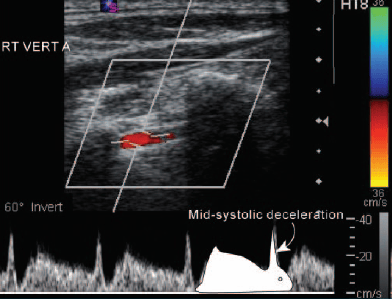
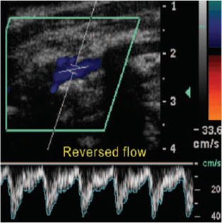
QUEM FAZ DOPPLER DE CARÓTIDAS AVALIA TAMBÉM AS ARTÉRIAS VERTEBRAIS.
NESTAS, TÃO IMPORTANTE QUANTO AVALIAR ESTENOSES É AVALIAR O PADRÃO DE FLUXO, QUE PODE NOS DAR PISTAS DE OCLUSÕES OU SUBOCLUSÕES DE ARTÉRIAS SUBCLÁVIAS OU DA INOMINADA…
ROUBO PARCIAL (FLUXO COM DESACELERAÇÃO MÉDIO-SISTÓLICA – SINAL DO COELHO):

ROUBO TOTAL (FLUXO REVERSO EM RELAÇÃO ÀS CARÓTIDAS):


DIÁSTOLE SEGUNDO OS EUROPEUS

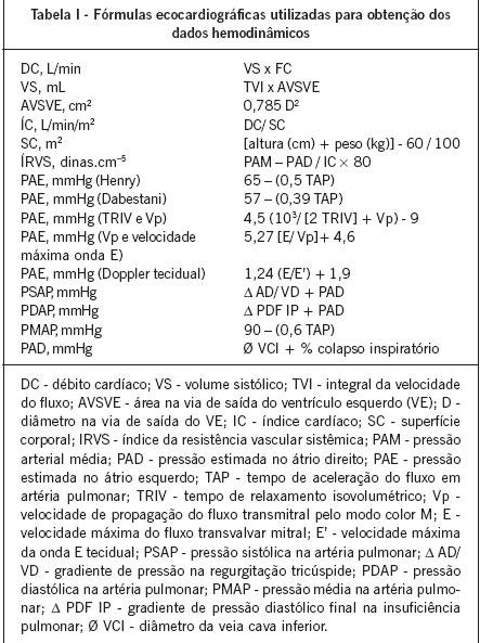
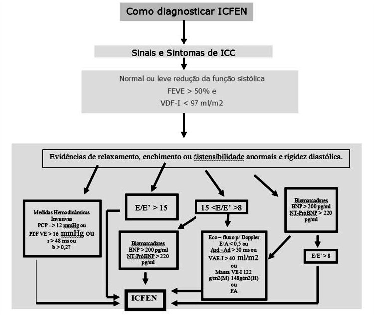
BELA REVISÃO DE DIASTOLOGIA, BASEADA NO GUIDELINE EUROPEU, PUBLICADA NA REVISTA DO DIC/DEPECO EM 2009:

http://departamentos.cardiol.br/sbc-depeco/v2/publicacoes/revista/2009/Revista03/09-doppler.pdf

XXXXXXXXXXXXXXXXXXXXXXXXXXXXXXXXXXXXXXXXXXXXXXXXXXXXXXXXXXX

REALMENTE BEM ELUCIDATIVA, CURTA, DIRETA E APLICÁVEL NA PRÁTICA DIÁRIA.

COMO JÁ DEFENDEMOS EM OUTRAS OCASIÕES, RELAÇÃO E/e´ E VOLUME ATRIAL TÊM DESTAQUE GARANTIDO NO ALGORÍTIMO.