CONCLUSION Currently, while 2D-SE is the standard examination for stress imaging in echocardiography, RT-3D-SE offers additional value to conventional 2D-SEin several areas. Three-dimensional imaging overcomes certain limitations of 2D imaging, but it remains limited by spatial and temporal resolution. While several studies have been published on the use of 3D-SE, larger studies are needed to provide more robust validation and prognostic information for 3D-SE. Technological advances in 3D imaging will further improve image quality and acquisition. With its ability to acquire the entire LV volume in a single beat, RT-3D-SE offers substantial promise for becoming the reference standard in the evaluation of patients with suspected CAD.
During dipyridamole stress echocardiography RT-3D imaging is highly feasible and shows a high concordance rate with standard 2D stress echo. 2D images take longer time to acquire and RT-3D is more time-consuming to analyze. At present, there is no clear clinical advantage justifying routine RT-3D stress echocardiography use.
The use of 3D echocardiography during SE reduces the time taken for image acquisition (58), improve foreshortened imaging planes (58) and increase reproducibility between imaging planes acquired at different stages of stress (59). New and relatively fast 3D ultrasound imaging devices are entering the market offering superior image quality, higher frame (3D volume) rates and ever-expanding capabilities that increase the potential for routine use.
Modes of 3D SE
3D systems offer several different imaging modes:
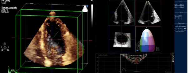
Real-Time 3D (RT3D) Multislice Imaging. Detection of haemodynamically-significant CAD with RT3D relies on the detection of changes in wall motion and thickening between rest and peak stress in the same way as 2D SE. The availability of multislice and multiplane RT3D permits simultaneous viewing of standard parasternal long, parasternal short and apical volumetric data, such that specificity and accuracy may be better than 2D SE (60). In fact, any chosen imaging plane of the LV can theoretically be visualised allowing a more detailed wall motion analysis than is currently available from standard 2D imaging planes. This improves detection of wall motion abnormalities in the apical segments and allows off-axis images to be interrogated, while preserving overall accuracy (60). Moreover, since the whole LV is imaged simultaneously, image acquisition becomes greatly simplified and faster (61).
Real-Time 3D Full Volume Data Acquisition (RT3DFV): With RT3DFV, full volume 3D datasets are acquired with the transducer positioned over the apex with the volume size adjusted to incorporate the entire left ventricle. Initial studies involving dobutamine stress echocardiography show shorter image acquisition times for 3D imaging than 2D imaging (62). There is no need to acquire parasternal images. As yet, data predominantly show equivalent overall accuracy compared to 2D SE with RT3DFV datasets (63).
3D volume data acquisition
The patient should be asked to stop breathing at end-expiration during the full volume acquisition to minimise issues with ‘fault lines’ between sub-volumes after reconstruction. Care should be given to ensure good-quality ECG gating and currently obtaining good-quality RT3DFV studies is difficult in patients with arrhythmias, particularly atrial fibrillation. With exercise SE, RT3DFV is more challenging and stitching artefacts due to hyperventilation and cardiac translation are more likely to occur, so much of the data acquired in existing publications relate to pharmacological stress.
In an otherwise suboptimal harmonic 3D SE study, LVO contrast has been shown to improve endocardial border delineation to a similar level to that achieved by contrast-enhanced 2D (64). A limitation to the use of LVO in 3D SE is that this does cause a reduction in temporal resolution (65). This has significance during peak exercise, when the heart rate is high and the number of 3D volumes per cardiac cycle is at levels that may be too low for diagnostic use.
Workflow and display
Present workflow issues for 3D stress analysis include cropping the 3D datasets to create a multiplane reconstruction equivalent to standard 2D image planes. However, this takes time and despite initially acquiring a 3D image, the time taken to create the necessary anatomical views may have an impact on the clinical workflow of SE. There are commercially available 3D stress software packages for processing 3D stress images that improve workflow, producing whole volume short axis slices for analysis. These derived 2D images can be ‘shuffled’ and displayed side by side for visual analysis in any format required. The key, however, to improving workflow for 3D SE will be the automation of the cropping and reconstruction process, and the quantification of LV wall motion and thickening.
There are two main techniques to achieve exercise stress – (a) semi-supine bicycle exercise or (b) a conventional treadmill test. Semi-supine bicycle is more sensitive than post-treadmill exercise (27). There are limited data to indicate similar sensitivity of SE during treadmill exercise to semi-supine bicycle but this is technically difficult (28). The aim of exercise SE is to achieve both the maximum target heart rate which is calculated (220 – age), although sensitivity of the test does not appear to fall providing imaging is performed above 85% of the maximum heart rate and maximal exercise. If a patient fails to achieve a minimum of 85% maximum heart rate, the rate of cardiovascular events in those who do not achieve their target but have normal images is higher than those who do achieve their target and have normal images (29). Similarly, failure to achieve a reasonable absolute level of exercise (defined as <7 metabolic equivalents (METs) for men and <5 METs for women) is a predictor of subsequent cardiac events even if SE is normal (30).