
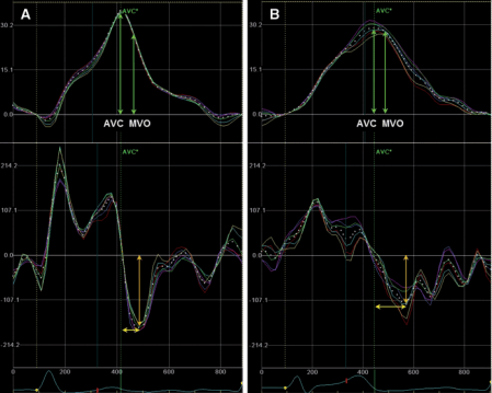
Measurements of LV rotation and back-rotation at 50 W of exercise. Representative cases of apical rotation curves (upper panel) and rotation velocity curves (lower panel) at 50 W exercise for Group 1 (A) and Group 2 (B). Apical back rotation at the MVO (%) was the proportion of the extent of recoiling from the peak apical rotation to the timing of the MVO. Minimal apical back-rotation velocity (degrees/s, orange arrow) and time-to-minimal apical back-rotation velocity after AVC (ms, yellow arrow) was defined from the rotational velocity vs. time curve. Apical back rotation at the MVO was greater in Case A compared with Case B. Minimal apical back-rotation velocity was lower and time-to-minimal apical back-rotation velocity after AVC was shorter in Case A compared with Case B. AVC, aortic valve closure; MVO, mitral valve opening.
Dynamic change in left ventricular apical back rotation: a marker of diastolic suction with exercise
https://doi.org/10.1093/ehjci/jex241
Conclusion
The lack of decrease in minimal LV pressure during exercise, a manifestation of impaired LV suction in early diastole, is linked closely with impaired LV apical back rotation during exercise. Dynamic changes in LV apical back rotation during exercise can be used as a non-invasive parameter of diastolic suction during exercise.