

.
Como transformar um consultório ou clínica em um bom negócio?
A maioria dos médicos não tem formação em gestão.
Acredita no ato médico como gerador de renda e pronto.
Isso funcionou muito bem antes dos convênios e a massificação da medicina.
Trabalho com um cardiologista de 78 anos que conta as histórias da antiga medicina, pré-convênios.
Agora está diferente. Como o ganho por ato médico é menor, foi necessário ampliar o número de atos por mês.
Aumento de volume de atendimento, tentando manter a qualidade. Mas qualidade custa tempo e dinheiro, que desperdiçados, tornam a empresa inviável.
Médicos isolados se uniram em clínicas para dividirem os custos, mas agem como se estivessem em seus consultórios separados.
Isso não ocorre em empresas, mesmo o português da padaria sabe que a empresa têm que ser lucrativa!
E parte do lucro têm que ser guardado para investimentos, ou no mínimo, manutenção da estrutura.
Recentemente me foi oferecida(para meu sócio Guilherme também) a gestão de 3 clínicas
Parecia um bom negócio, até eu conhecer de fato as estruturas.
O dono havia retirado todo o lucro nos últimos 5 anos, a empresa estava sucateada!
Funcionários sem treinamento adequado, computadores com defasagem de 10 anos, manutenção ausente, aparelhos velhos e necessidade urgente de uma reforma.
A empresa gerava 15 mil de lucro mensal, mas precisava de pelo menos 10 mil por mês em investimentos para voltar a ser um bom negócio.
Como uma empresa que faz 500 ecos por mês foi parar nesse buraco?
A resposta ajudaria centenas de clínicas pelo país, mergulhadas na ausência de gestão administrativa.
Mês: agosto 2008
FDA em crise. Quem vigiará as grandes empresas

Sen Arlen Specter (R, Pa) said the White House is delaying the swift provision of an additional $275 million to the US Food and Drug Administration (FDA) by requesting that the funds be included in the fiscal year 2009 budget rather than the FDA’s current budget. The money is intended to help the agency protect the nation’s food supply, ensure safer drugs, and modernize its facilities.
Specter said he had worked to get the FDA an additional $275 million in the Senate version of the fiscal year 2008 supplemental appropriations bill, which, if approved, would provide the funds to the agency no later than September 30. In a June 10 letter to Michael Leavitt, secretary of the Department of Health and Human Services, Specter wrote that the Bush administration’s request is “drastically hindering necessary immediate relief” because the additional funding would not be provided until March or April 2009.
“The 81 deaths due to contaminated heparin and the 1 suspected death in the ongoing Salmonella outbreak show that we cannot wait 9 months to give the FDA the resources needed to protect the public,” Specter wrote.
Mike Mitka
Dissecção da aorta vista por dois métodos complementares.
Panel A. Aortic dissection with a systolic flow in the true lumen (TL) of ascending aorta on TEE. A continual flow can be seen in the false lumen (FL) in which an intraluminal thrombus (T) is evident.

Panel B. A hypermobile double lobar thrombus connected to the penetration canal (P) heading to left atrial roof.
Panel C. Continuous turbulent jet heading from the aorto-left atrium fistula canal (P) to left atrial roof.
Panel D. Double intimal tear in descending aorta.
Panel E. A reconstructed three-dimensional CT image using volumetric rendering method. An evident aneurysmatic dissection of ascending aorta with a rupture of aortic adventitia and penetration into left atrium (arrow).
Panel F. An extensive thoracic aorta aneurysm dissection penetrating into left atrium. A tricuspid aortic valve (Ao), left ventricle (LV), and an intimal line (I) separating true aortic lumen (TL) from false lumen (FL) can also be noticed. Hypodense masses with an irregular margin lining the edges of a proximal part of a false lumen are thrombi (T) penetrating into the left atrium (LA).
Panel G. Short axis CT image of ascending and descending aorta.
Panel H. Distal part of dissection in area of right common iliac artery (white arrow).
Panel I. A view of a false lumen with a noticeable penetration opening into left atrium. The arrows are pointing towards its CT angiography and TEE correlates. The true aortic lumen is compressed by a suction tube.
BLOG ECHOTALK: MAIS DE 40 MIL VISITAS

No último mês (Fonte Google Analytcs):
4623 Visitas
1280 visitantes diferentes.
2min 44s Tempo médio da visita
1287 Visitantes que repetem a visita
Lealdade do visitante
4 vezes 82
5 vezes: 59 leitores
6 vezes: 45 leitores
7 vezes: 37 leitores
8 vezes: 31 leitores
9-14 vezes: 124 leitores
15-25 vezes: 138 leitores
26-50 vezes: 131 leitores
51-100 vezes: 80 leitores
101-200 vezes: 104 leitores
201+ vezes: 140 leitores
Para um blog de assunto específico, um sucesso!
Obrigado
Microbolhas? Continua o mistério…
.
Quer comprar Definity?
Está difícil.
Nenhum sinal da Cristália, atual vendedora de microbolhas.
Imagina se eu tivesse um serviço que dependesse do fornecimento inenterrupto do contraste.
Nos EUA, está a venda normalmente Aqui
DIC PARA O DEPECO

.
No blog foi assim:
72 votantes
36 Contra o DIC
29 A favor do DIC
4 Não se importam
3 Não resistem a nossa presidente!
E não se fala mais nisso!
Complicado mas fascinante.
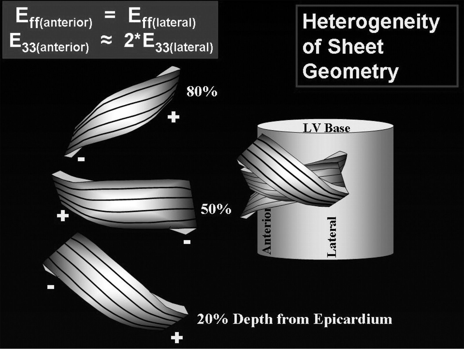
Fiber and sheet orientations. A model synthesizing fiber and sheet data from anterior and lateral equatorial regions of the heart. The illustration on the left represent sheets at 20%, 50%, and 80% wall depth from the epicardium. Black lines on the surface of the sheets represent measured {alpha} qualitatively, with negative {alpha} values near the epicardium, values of 0° at midwall, and positive {alpha} values near the epicardium. Left margins of the sheets represent data from the anterior wall, with sheets belonging to the negative β family at 20%, to the positive β family at 50%, and to the negative β family at 80% wall depth. Right region of the sheets represent data from the lateral region, with sheets belonging to the positive β family at 20%, to the negative β family at 50%, and to the positive β family at 80% wall depth from the epicardium.
.Circulation. 2008;118:713-721
Heterogeneity of Left Ventricular Wall Thickening Mechanisms
Tendão anômalo é igual cavalo bravo, louco pra te derrubar…

.
Conversando com um colega cardiologista, ele me disse que o eco havia falhado em reconhecer uma hipertrofia septal assimétrica.
Tinha inclusive ECG de repouso alterado e ergométrico positivo com 5 mm em paciente hígido de 35 anos.
Ecocardiograma normal com ressonância mostrando hipertrofia septal.
Pedi para ele me enviar o paciente, sem custos, e refiz o exame.
Resultado:
Tendão anômalo com inserção no septo basal, dando a falsa impressão de hipertrofia.
Tendôes assim podem conter sistema de condução e alterar o ECG.
Fiz várias fotos, com zoom, sem zoom, mostrei para um colega na hora.
O colega cardiologista ficou decepcionado, acredita tanto na ressonância…
Mais fotos em breve.
FUTOROLOGIA OU EXAME DE CARÓTIDAS?
.
Paciente de risco intermediário realizou exames de carótidas com os achados acima.
Espessamento difuso da íntima-média e fluxo na interna indicativo de aterosclerose intra-craniana aos 65 anos!
Dois fortes marcadores de eventos desfavoráveis em um paciente de risco moderado.
Para uma parte dos cardiologistas, isso o coloca no grupo de alto risco e merecedor de metas de atividade física, dieta, LDL, HDL e triglicérides mais rígidas.