

http://zaxpie475.blogspot.com/2008/12/tudo.html
.
Veja na filmagem acima a “sala” de ecocardiografia em funcionamento.
Realiza 200 exames por mês.
Em pequenos espaços, vive a ecocardiografia atual.
Em 2009, você não vai montar a sua sala?!?!?
.
BlogBlogs.Com.Br
Dia: 3 de dezembro de 2008
Tomo é boa, mas ainda não está pronta.
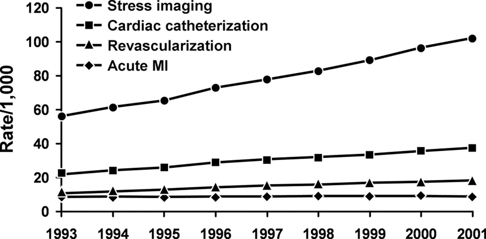
Noninvasive Diagnosis and Prognosis Assessment in Chronic Coronary Artery Disease
Stress Testing With and Without Imaging Perspective
Raymond J. Gibbons, MD
From the Division of Cardiovascular Diseases and Internal Medicine, Mayo Clinic, Rochester, Minn.
.Conclusão:
O uso do teste de esforço com ou sem imagem não invasivos no diagnóstico e prognóstico da avaliação DAC crônica está bem estabelecido. É enfatizado o seu importante papel na prática clínica existente diretrizes e apoiado por uma extensa base de dados. CT angiografia coronária é uma técnica muito promissora. Os avanços técnicos nesta matéria, no seio dos últimos anos são verdadeiramente espantosos. No entanto, muito mais pesquisas são necessárias antes que esta técnica pode ser aplicada sobre uma ampla base na prática clínica. Entretanto, a abordagem preferida deve continuar a ser teste ergométrico com ou sem imagem. Esperemos que, futuramente irá proporcionar estudos mais provas para apoiar o papel da CT angiografia em pacientes adequados e demonstrar que o seu benefício justifica o investimento adicional necessário em pessoal, formação e equipamento.
Three-Dimensional Transesophageal Echocardiography in a Patient With Early Failure of Mitral Valve Repair
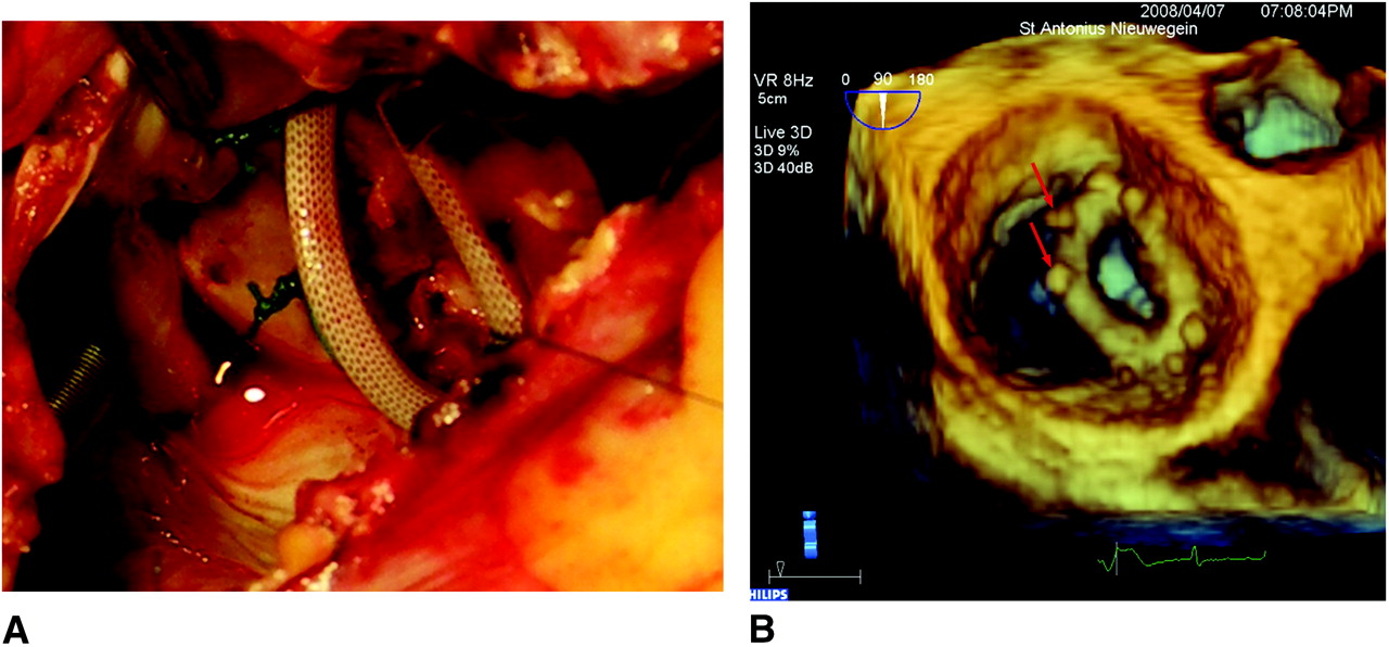
Figure 2. A, Peroperative photograph confirming partial dehiscence of the mitral valve annuloplasty ring. Similarity to the preoperative 3D images is excellent (B; Movie 1). Notice the torn stitches that are still connected to the annuloplasty ring. These are also visible on the 3D images (red arrows).
Three-Dimensional Transesophageal Echocardiography in a Patient With Early Failure of Mitral Valve Repair
Why Are We Still Looking at a Three-Dimensional Structure in 2 Dimensions?
.
Por que ainda olhamos de maneira bi-dimensional uma estrutura tri-dimensional.
Bela pergunta a ser respondida.
COARCTAÇÃO DA AORTA

Recommendations for Interventional and Surgical Treatment of Coarctation of the Aorta in Adults
Class I
1. Intervention for coarctation is recommended in the following circumstances:
a. Peak-to-peak coarctation gradient greater than or equal to 20 mm Hg. (Level of Evidence: C)
b. Peak-to-peak coarctation gradient less than 20 mm Hg in the presence of anatomic imaging evidence of significant coarctation with radiological evidence of significant collateral flow. (Level of Evidence: C)
2. Choice of percutaneous catheter intervention versus surgical repair of native discrete coarctation should be determined by consultation with a team of ACHD cardiologists, interventionalists, and surgeons at an ACHD center. (Level of Evidence: C)
3. Percutaneous catheter intervention is indicated for recurrent, discrete coarctation and a peak-to-peak gradient of at least 20 mm Hg. (Level of Evidence: B)
4. Surgeons with training and expertise in CHD should perform operations for previously repaired coarctation and the following indications:
a. Long recoarctation segment. (Level of Evidence: B)
b. Concomitant hypoplasia of the aortic arch. (Level of Evidence: B)
ACC/AHA 2008 Guidelines for the Management of Adults With Congenital Heart Disease: Executive Summary: A Report of the American College of Cardiology/American Heart Association Task Force on Practice Guidelines (Writing Committee to Develop Guidelines for the Management of Adults With Congenital Heart Disease)
. A partir de 20 mmhg na coarctação já seria indicativo de intervenção, pela diretriz fresquinha.
Ao Ecocardiograma, devo crer.
Usando a equação de Bernolli simplificada ou com a inclusão da Velocidade pré estenose…
ANEURISMA GIGANTE DO APÊNDICE ATRIAL
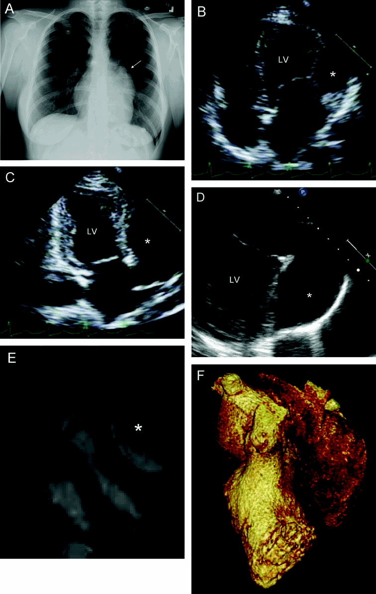
cIRCULATION Volume 118(23), 2 December 2008, pp 2393-2394
Novo portal do DEPECO

.
O novo site do DEPECO está moderno, bonito e funcional!
Vamos visitar e dar sugestões, mudanças para melhor serão sempre bem-vindas.