
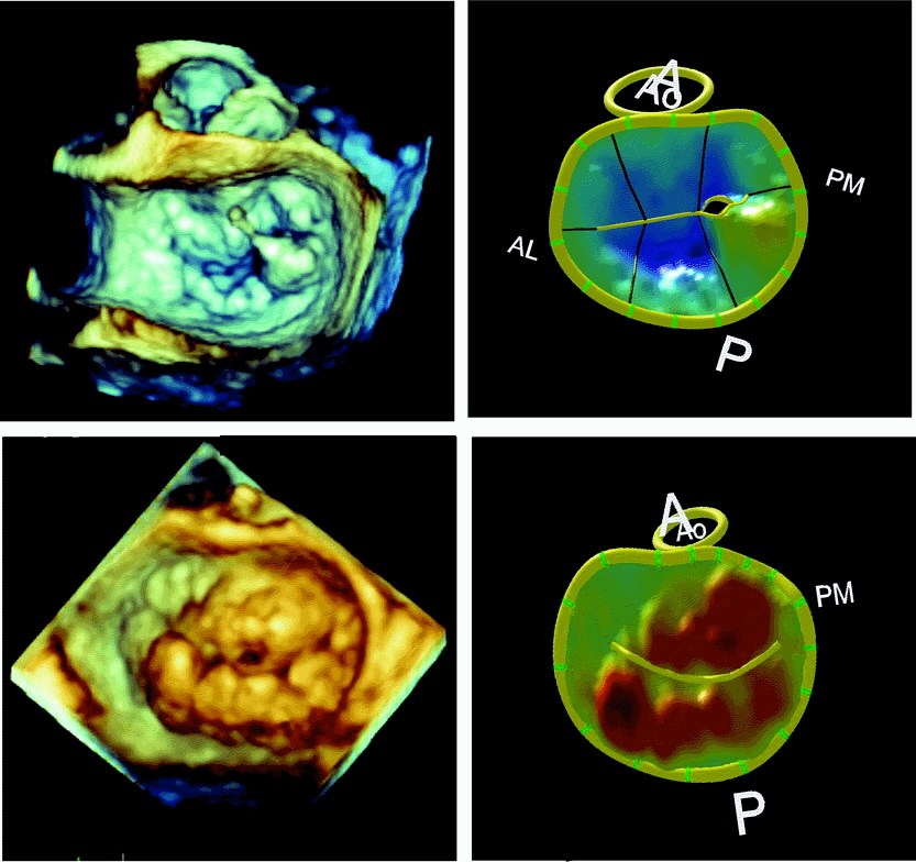
Você está pronto para o 3D de rotina?

Real-Time 3-Dimensional Echocardiography: An Integral Component of the Routine Echocardiographic Examination in Adult Patients?
Circulation Volume 119(2), 20 January 2009, pp 314-329
.
“Currently, many laboratories perform a complete 2DE study, followed by a focused 3D examination, in patients with specific pathologies in which RT3DE imaging could potentially provide additional diagnostic information. It can be anticipated that a full-volume acquisition of the left ventricle will be performed in every patient to obtain LV volumes and EF. The 3D images should be stored in a digital archiving system with the 2D study to allow integrated interpretation of all images and incorporation of 3D findings into the report.

Future advances in transducer and computer technology will result in several important improvements that will further enhance the clinical application of RT3DE imaging. One highly desirable improvement is the ability to acquire wider-angle pyramid of data with and without color flow in a single cardiac cycle. This will shorten data acquisition and eliminate stitching artifacts. Furthermore, future improvements in both spatial resolution and temporal resolution of the transthoracic RT3DE imaging, which are still below those of 2DE, will broaden the spectrum of patients who can be imaged with this modality. Further miniaturization of the 3D MTEE technology will allow 3D TEE imaging in pediatric patients and the development of real-time 3D imaging intracardiac catheters. Future software developments will allow new types of sophisticated quantitative analysis of the cardiovascular anatomy and function, including the fusion of RT3DE data with other 3D imaging modalities such as magnetic resonance and computed tomography.”
.
A pergunta não é se o 3D já está pronto, e sim se nós estamos prontos para o 3D.
Insisto que usar o 3D após o exame em 2D é um erro, e prejudica a ampla diusão do método.
É para usar o 3D do início ao fim.
Sempre ele, o volume atrial.


Left atrial remodelling in patients with myocardial infarction complicated by heart failure, left ventricular dysfunction, or both: the VALIANT Echo Study
European heart journal Volume 30(1), January 2009, pp 56-65
.
Aims: To assess the relationship between left atrial (LA) size and outcome after high-risk myocardial infarction (MI) and to study dynamic changes in LA size during long-term follow-up.

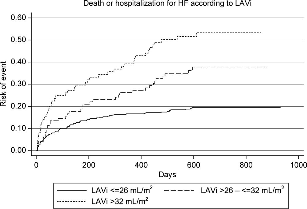
Methods and results: The VALIANT Echocardiography study prospectively enrolled 610 patients with left ventricular (LV) dysfunction, heart failure (HF), or both following MI. We assessed LA volume indexed to body surface area (LAVi) at baseline, 1 month, and 20 months after MI. Baseline LAVi was an independent predictor of all-cause death or HF hospitalization (P=0.004). In patients who survived to 20 months, LAVi increased a mean of 3.00 ± 7.08 mL/m2 from baseline. Hypertension, lower estimated glomerular filtration rate, and LV mass were the only baseline independent predictors of LA remodelling. Changes in LA size were related to worsening in MR and increasing in LV volumes. LA enlargement during the first month was significantly greater in patients who subsequently died or were hospitalized for HF than in patients without events.

Conclusion: Baseline LA size is an independent predictor of death or HF hospitalization following high-risk MI. Moreover, LA remodelling during the first month after infarction is associated with adverse outcome.
.
O volume atrial é o melhor marcador de disfunção diastólica e um ótimo índice de prognóstico.
Nesse artigo, fica evidente que é um marcador precoce de má evolução.

Eu vejo Aterosclerose todos os dias.

.
Não sei quem compartilha comigo essa perseguição diária ao diagnóstico de Aterosclerose, mas mesmo imagens pouco alteradas, como a abaixo, deixam um examinador como eu maravilhado.
O paciente têm 80 anos!
Quando vejo a Aterosclerose desse idoso, entendo melhor a do paciente de 50 anos, entendo as diferenças, não pelo que vejo, mas pelo que não vejo!
.
“O médico deve falar do que é invisível. O que é visível, pertence ao seu conhecimento, como a qualquer outro que não seja médico e reconheça a doença pelo sintoma. Mas isto está longe de fazê-lo médico: torna-se médico quando, e somente quando, souber o invisível e imaterial, eficazes”

Paracelso, 1528.

Dica de livro de Eco


.
Edição de 2007 de um livro realmente bom, tendo o famoso Jamil Tajik como co-autor.
Um pouco caro, mas são 431 páginas, não é um manual qualquer.
Li a edição anterior e encomendei essa.

Pesquisa de Doppler do Anel Mitral


.
Clinical Utility of Doppler Echocardiography and Tissue Doppler Imaging in the Estimation of Left Ventricular Filling Pressures

A Comparative Simultaneous Doppler-Catheterization Study

S. R. Ommen, MD; R. A. Nishimura, MD; C. P. Appleton, MD; F. A. Miller, MD; J. K. Oh, MD; M. M. Redfield, MD; A. J. Tajik, MD
.
Tissue Doppler Measurements
TDI of the mitral annulus was obtained from the apical 4-chamber view. A 1.5-mm sample volume was placed sequentially at the lateral and medial mitral annulus. Analysis (Figure 1) was performed for the early (E’) and late diastolic velocity (A’): peak velocity (E’, A’), time to peak velocity (E’at, A’at), DT (E’DT, A’DT), and duration of velocity profile (E’dur, A’dur). These variables were analyzed individually, as the average of the medial and lateral annulus, and as the maximum of the medial and lateral annulus.
.
O estudo fundamental de Doppler do anel, disponível acima, e tendo o famoso Nishimura como co-autor, manda fazer em duas posições, lateral e septal.
.
Só 27% dos leitores seguem a recomendação mais aceita.
Metade escolhe um lado e pronto.
Eu votei no volume atrial, mais importante para definir disfunção diastólica significativa.
Quando quero acreditar no Doppler do anel, faço dos dois lados.

Echotalk = Escola de ex-alunos de São Paulo


.
É sério, dos 6 alunos matriculados para Fevereiro e Março, para cursos individuais, 5 são ex-alunos de uma grande escola de São Paulo!!!
Só 1 não tentou aprender em Powerpoint, e veio direto pra Echotalk.
Isso é que é marketing eficiente, sucesso de vendas com “ligeira” falha em entregar o que promete…

Novas idéias sobre a Diástole


.Titin alters cardiomyocyte stiffness through isoform shifts, phosphorylation, and titin–actin interaction. A, Sarcomeric structure with detailed view of I-band region of N2B titin isoform showing tandem immunoglobulin (Ig), N2B, and elastic PEVK segments. B through D, Shift from N2B to N2BA titin isoform (B) and phosphorylation by PKG or PKA at S469 (C) reduce stiffness of the elastic PEVK segment, whereas wider titin–actin overlap at the Z-disc possibly increases stiffness of the elastic PEVK segment (D).
From: Borbely: Circ Res, Volume 104(1).January 2, 2009.12-14
.
Um conceito bem firmado era a participação da matriz extracelular nas alterações crônicas que levam a disfunção diastólica.
Alterações agudas eram dadas como relacionadas a isquemia, na maioria das vezes.
Estudo experimental demonstrou alteração aguda da diástole com intervenção química na matriz extracelular.
.
A divisão de disfunção ventricular diastólica em crônica ou aguda ficou menos visível.
Mais um exemplo da dinâmica absoluta da fisiologia cardíaca, com a coronária instável desde a adolescência, bem como a diástole pronta para ser alterada a cada momento.

ECO DE REPOUSO, PARA OCLUSÃO.


ARTIGO
Transthoracic echocardiography in the detection of chronic total coronary artery occlusion
.
Fico surpreso com as descobertas para o ecocardio de repouso!
A melhora das máquinas, finalmente está revertendo em melhora do método.

Tema livre merece respeito


.
Apresentei meu primeiro tema livre em 98, no congresso DEPECO. Foi o primeiro da ecocardiografia da UNICAMP, depois apresentei na SOCESP e Congresso Brasileiro.
Acho importante para a instituição aparecer nesses eventos.
Mas é preciso ter paciência!
Colocam você em cada buraco, cada sala escondida, sem horário para os painéis, uma confusão…
Ainda valorizo muito esse tipo de apresentação, mostra que estamos pensando no assunto, não é só trabalho de rotina. Qualquer laboratório de ecocardiografia pode produzir um tema.
Uma passagem de 2002 me ensinou muito:
Estava esperando há mais de 1 hora para apresentar meu tema em painel, marcado para 10 h, as 11:30 não havia começado!
Decidi sair sem apresentar! Enquanto retirava os grampos, olhei para o lado e vi o Prof. Antonio Carlos Pereira Barreto, sentado calmamente, esperando para apresentar seu painel.
Como ele aguentava esperar?
Simples, ele estava ali por ele e seus colaboradores, não pelos figurões da cardiologia…
Sentei e esperei pela comissão de apresentação.

A dança do coração


Cardiac Mechanics Revisited: The Relationship of Cardiac Architecture to Ventricular Function
Circulation 2008;118;2571-2587
.
Note os movimentos dos vetores em diferentes regiões do coração, alguns em sentido horário, outros anti-horário.
Essa figura vales por 10 textos de Strain.
Veja o artigo completo