http://journal.frontiersin.org/Journal/10.3389/fphys.2012.00090/full
.

.
During Exercise
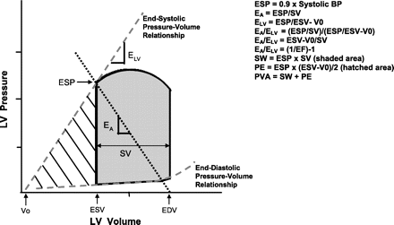
During exercise EA has been shown to increase (Najjar et al., 2004; Otsuki et al., 2006), decline (Asanoi et al., 1992), or remain unchanged (Cohen-Solal et al., 1998). The response of EA during exercise is dependent on the changes in its components and has important consequences on the Frank–Starling mechanism (Chantler et al., 2011). EA is linearly related to heart rate and peripheral resistance, and inversely related to compliance (Segers et al., 2002; Chemla et al., 2003, Otsuki et al., 2006; Chantler et al., 2011). Both resistance and compliance usually decrease during exercise (reflecting less resistance to blood flow in the microcirculation, but increased stiffness of the conduit arteries) and the relative contribution of the pulsatile component (compliance) to EA increases, so that by 80% of peak exercise the resistive and the pulsatile components provide nearly equal contributions to EA (Otsuki et al., 2006). With advancing age, the ability to increase heart rate, and lower resistance during exercise is blunted.
.
Envelhecimento e sua relação com a diástole passa pela resistência periférica. Está aqui a explicação para os falso doentes da diástole e verdadeiros doentes da resistência periférica?
.