
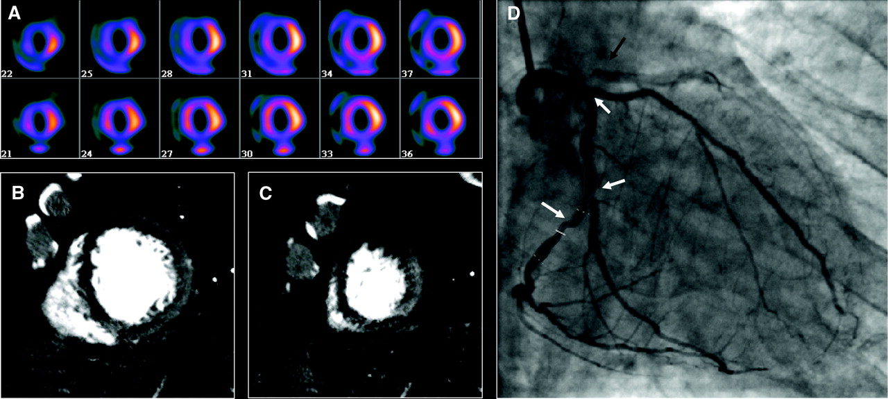
Images from 64-row detector CTP. A, Partially reversible perfusion deficit in the territory of the LAD and a primarily fixed perfusion deficit in the inferior wall on radionuclide myocardial perfusion imaging with increased subdiaphragmatic tracer uptake (stress, upper panels; rest, lower panels). B and C, Adenosine stress CTP shows a dense perfusion deficit in the LAD territory, as well as a subendocardial perfusion deficit in the inferior and lateral walls. D, Invasive coronary angiogram demonstrates a left-dominant system with a totally occluded LAD (black arrow) as well as intermediate and high-grade stenoses in a large ramus intermedius, the body of the left circumflex, an the ostium of the obtuse marginal artery (white arrows).
Artigo
Adenosine Stress 64- and 256-Row Detector Computed Tomography Angiography and Perfusion Imaging
A Pilot Study Evaluating the Transmural Extent of Perfusion Abnormalities to Predict Atherosclerosis Causing Myocardial Ischemia
Cardiovascular Imaging. 2009;2:174-182
.
Conclusions— The combination of CTA and CTP can detect atherosclerosis causing perfusion abnormalities when compared with the combination of quantitative coronary angiography and SPECT.