

.
Outro dia um aluno perguntou se eu conhecia algum site com exames “diferentes” de ecocardiografia!!!.
.
Serve esse? rs
.
Mês: junho 2010
Rejeição ao coração transplantado
.
Paciente jovem, transplante há 6 meses com dispnéia e fraqueza há 3 dias.
Exame há dois meses com cavidades e ejeção normais.
.
Ecocardiografia e Buenos Aires, uma bela combinação!

.
Aqui.
Destaques:
12:00 a 12:45 Salón Gaudí Sesión Plenaria 3: Evaluación de la función ventricular derecha Dr. Roberto Lang
12:00 a 12:45 Salón Gaudí Sesión Plenaria 1 La Utilidad del Eco 3D en la Evaluación de la Valvulopatía Mitral. Dr. R. Lang
16:45:17:00 Cuantificación de la Estenosis Aórtica. ¿Puede el eco 3D proveer valor agregado? Dr. R. Lang
17:45 a 18:30 Salón Gaudí Sesión Plenaria 2 Errores y limitaciones de la Ecocardiografía en la cuantificación de la Estenosis Aórtica Dr. Arturo Evangelista
.
Congresso Mundial : Edema pulmonar
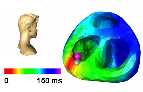
The Role of Cardiac Dyssynchrony in the Pathogenesis of Acute
Hypertensive Pulmonary Edema
Andrei-Dumitru Margulescu1,, Roxana Cristina Sisu1,2, Maria Florescu2, Mircea
Cinteza1,2, Dragos Vinereanu
.
Introduction: The role of acute cardiac dyssynchrony during acute hypertensive pulmonary edema (AHPE) has not been evaluated. Methods: 51 consecutive patients (69 11 years, 31 women), admitted to an intensive cardiac care unit with the diagnosis of AHPE, were evaluated by conventional and tissue Doppler echocardiography, during the acute episode, and again at 48 to 92 hours. Inclusion criteria were: acute respiratory distress within the preceding 8 hours with clinical and radiological pulmonary congestion, systolic BP 160 mmHg, and sinus rhythm; patients with acute myocardial infarction or moderate and severe left-sided valvar diseases were excluded. Inter-ventricular dyssynchrony was assessed from aorto-pulmonary
flow delay (APD), and maximal difference between peak-to-peak myocardial velocity tracings of any LV segment and the RV segment (Inter). Intra-ventricular dyssynchrony was assessed from maximal delay of peak-to-peak systolic myocardial velocities (MxS) and peak systolic displacement (MxD) in 6 basal segments of opposing LV walls, standard deviation of time-to-peak myocardial systolic velocity in 12 myocardial segments (Yu index), and aortic pre-ejection time (APT). Atrio-ventricular (AV) synchrony was assessed from diastolic filling time (% of the cardiac cycle,%DFT). Severity and mechanisms of secondary mitral regurgitation (MR) were assessed using qualitative (visual grading by a scale from 1 to 4), and quantitative
measurements (vena contracta, MR area, tenting area of the mitral valve, and diameter of the mitral annulus).
Results: Complete left bundle branch block (LBBB) was present in 12 patients during ACPE and in 13 at follow-up (p 0.5). Inter-ventricular (APD: 26 vs. 25 ms, p 0.88; Inter: 45 vs. 54 ms; p 0.36) and intra-ventricular dyssynchrony indexes (MxS: 42 vs. 61 ms,p 0.12; MxD: 28 vs. 29 ms, p 0.44; Yu index: 35 vs. 37 ms, p 0.63; APT: 90 vs. 89 ms, p 0.82) showed that AHPE was not associated with acute dyssynchrony, after adjustment for differences in heart rate (94 vs. 75 bpm, p 0.001).%DFT was reduced during AHPE compared with follow-up (40% vs. 48%, p 0.001), suggesting impaired AV synchrony induced by the increased HR. Duration of the A wave did not differ between evaluations (138 ms vs. 140 ms, p 0.7). The severity of MR was similar between evaluations.
Conclusion: Impaired atrio-ventricular synchrony, but not acute inter- and intra- ventricular dyssynchrony are involved in the pathogenesis of AHPE.
3D ou pirâmide?
.
Outro dia, uma leitora perguntou do formato do 3D. Expliquei, mas esse filme é bem melhor!!!
.

Obstrução do seio coronário

Coronary Sinus Obstruction by Primary Cardiac Lymphoma as a Cause of Dyspnea Due to Significant Diastolic Dysfunction and Elevated Filling Pressures
(J Am Soc Echoc 2010;23:682.e5-682.e7.)
.
Caso raro de obstrução do seio coronário por Linfoma com dilatação gigantesca do seio.
.
Indicado por Leandro Serafim.
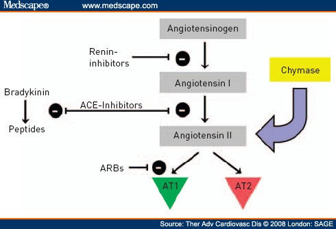
O inferno dos BRA.
.
A guerra comercial travada entre os bloqueadores do receptor da angiotensina vai ter uma breve pausa para defesa.
.
Estudos apontando aumento de neoplasia de pulmão (Modest lung-cancer signal with angiotensin-receptor blockers).
“Dr Steven E Nissen (Cleveland Clinic, OH) calls the meta-analysis “disturbing and provocative, raising crucial safety questions for practitioners and the regulatory community.”
.
E ainda mais, o FDA vai revisar os estudos ROADMAP E ORIENT, acusados de aumentarem o risco de morte em diabéticos.
.
Principalmente depois do ON TARGET, o futuro dos BRA não parece tão promissor.
.
Quase todo trombo têm uma fonte.
.
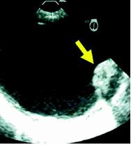
Complex Plaques in the Proximal Descending Aorta
.
An Underestimated Embolic Source of Stroke
.
Stroke. 2010;41:1145
.
Potential embolization from DAo plaques was then identified in 19 of 57 patients (33.3%) with determined and in 9 of 37 patients (24.3%) with cryptogenic stroke.
Conclusions— Retrograde flow from complex DAo plaques was frequent in both determined and cryptogenic stroke and could explain embolism to all brain territories. These findings suggest that complex DAo plaques should be considered a new source of stroke.
.
Mais um achado interessante do renascido transesofágico, placas na aorta descendente como fonte de trombos para AVC.
.
A pesquisa de fontes emboligênicas no AVC, pedido comum na ecocardiografia, precisa mesmo ser feita com mais empenho.
.
Quase todo sopro têm explicação.
.
Sexo feminio 66 anos em exame de rotina, assintomática.
.
Enviado por Leandro Serafim.