
Recurrent Pericardial Constriction Circulation. 2008;118:1685-1688
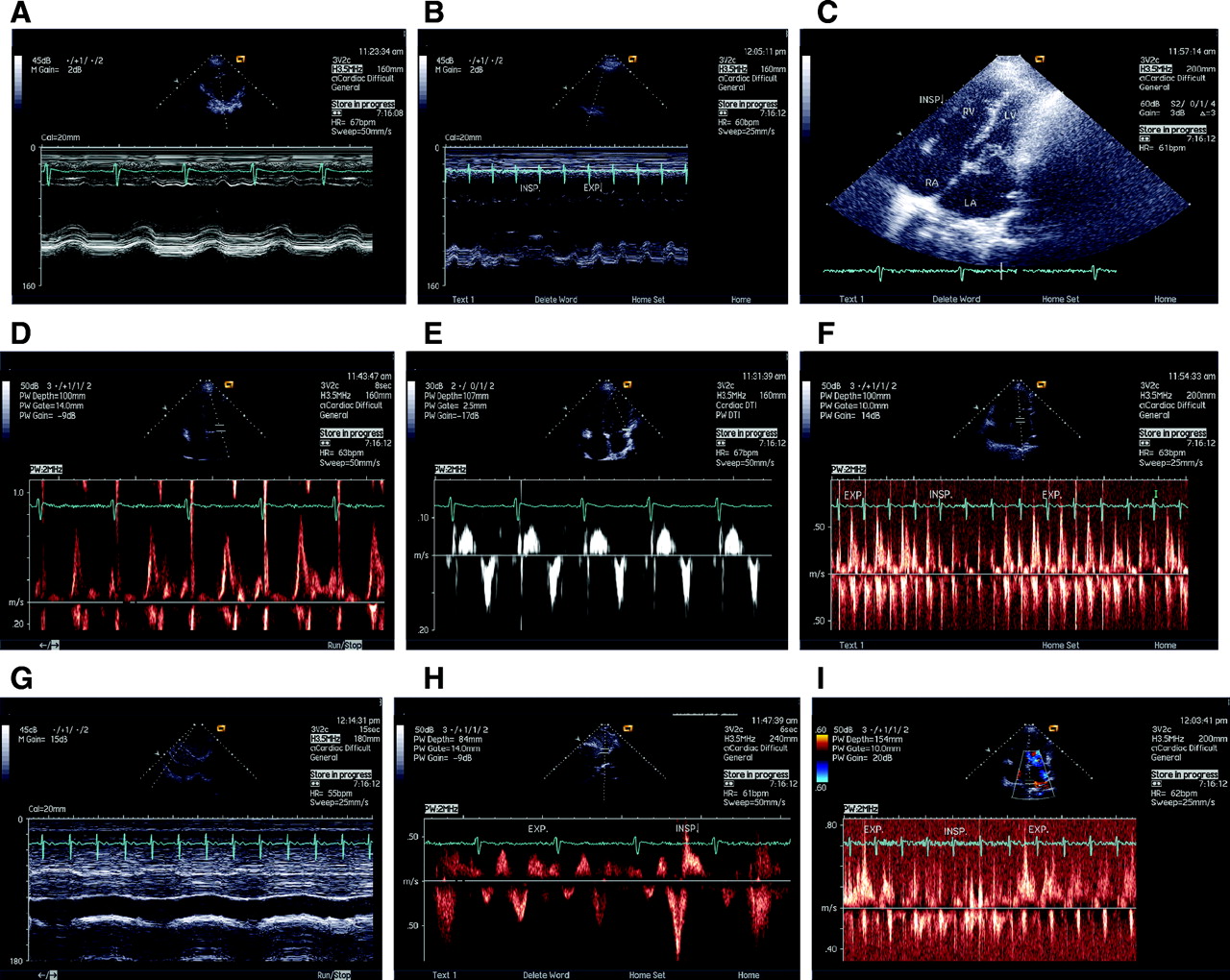
Echocardiographic findings. A, M-mode with abnormal-flat septal motion and thick hyperechogenic pericardium. B, M-mode with septal bounce with inspiratory displacement of the septum toward the left ventricle. C, Apical 4-chamber view during inspirium with the ventricular septum displaced to the left. D, Restrictive left ventricular filling profile with large E-wave and short deceleration time. E, Preserved mitral annular tissue Doppler early diastolic velocity (Ea). F, Exaggerated increase of left ventricular filling velocities on experium. G, Dilated inferior vena cava without respiratory change of diameter. H, Inspiratory increase of velocities through the hepatic veins. I, Exaggerated increase in pulmonary venous velocities.