

Natural History of Very Severe Aortic Stenosis
Circulation. 2010;121:151-156.
.
Methods and Results— We prospectively followed 116 consecutive asymptomatic patients (57 women; age, 67±16 years) with very severe isolated aortic stenosis defined by a peak aortic jet velocity (AV-Vel) ≥5.0 m/s (average AV-Vel, 5.37±0.35 m/s; valve area, 0.63±0.12 cm2)
During a median follow-up of 41 months (interquartile range, 26 to 63 months), 96 events occurred (indication for aortic valve replacement, 90; cardiac deaths, 6). Event-free survival was 64%, 36%, 25%, 12%, and 3% at 1, 2, 3, 4, and 6 years, respectively. AV-Vel but not aortic valve area was shown to independently affect event-free survival. Patients with an AV-Vel ≥5.5 m/s had an event-free survival of 44%, 25%, 11%, and 4% at 1, 2, 3, and 4 years, respectively, compared with 76%, 43%, 33%, and 17% for patients with an AV-Vel between 5.0 and 5.5 m/s (PII) than those with an AV-Vel between 5.0 and 5.5 m/s (27%; P=0.03).
Conclusions— Despite being asymptomatic, patients with very severe aortic stenosis have a poor prognosis with a high event rate and a risk of rapid functional deterioration. Early elective valve replacement surgery should therefore be considered in these patient
.
Uma velocidade acima de 5m/s sugere uma pressão intraventricular de 220 mmhg para uma sistólica de 120 mmhg.
É muita pressão a ser gerada por um músculo cardíaco a cada batimento.
Normalmente o ecocardiograma não indica cirurgia isoladamente, mas nesse caso parece haver justificativa.
.
Mês: janeiro 2010
URGENTE: Nova tabela para medidas no modo M anatômico.

.
Faço em todos os casos.
Meço as paredes e cavidades sem o modo M anatômico e depois, lado a lado (foto), faço com o modo M anatômico.
As medidas são significativamente menores.
Isso têm sido notado por outros colegas.
.
Um método diagnóstico só é bom quando possui uma tabela de normalidade confiável, o que já não acontece mais nas medidas cavitárias ao modo M.
.
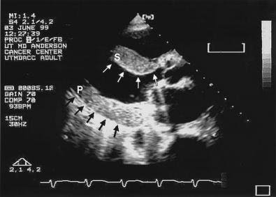
Amiloidose
.
Chama a atenção para o espessamento das valvas!
.
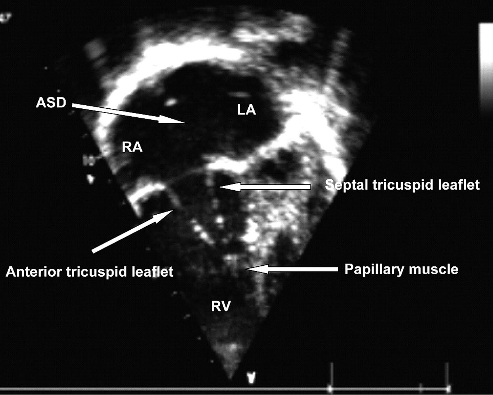
Endocardite de septo interatrial.

A 24-year-old man presented with a short history of fever and swollen lower limbs. Physical examination revealed signs of significant mitral regurgitation and blood cultures grew Streptococcus viridans. Transoesophageal and transthoracic echocardiography demonstrated prolapse of the mid-portion (P2) of the posterior mitral valve leaflet with severe mitral regurgitation. There was a 9mm by 9mm vegetation attached to the abnormal leaflet . Echocardiography also identified a 10mm secundum atrial septal defect with left to right shunting of the mitral regurgitation jet through the defect . Along the inferior left atrial side of the septal defect were multiple small mobile masses in keeping with endocarditis of the atrial septum . The patient was treated with a four-week course of antibiotics with rapid clinical improvement and resolution of the vegetations.
.
Insuficiência aórtica e PHT.

Thirty four patients with aortic regurgitation were studied by continuous wave Doppler ultrasound. In 30 of these the regurgitation was graded by cineangiography as mild, moderate, or severe and in four severe regurgitation was confirmed at operation. The half times of the aortoventricular pressure differences obtained with Doppler compared well with those obtained from pressure recordings at catheterisation. The relation between pressure half times and cineangiographic gradings of severity was not consistent. Similarly, a control group of patients without aortic regurgitation showed a wide range of the invasively recorded pressure half times which overlapped with those in patients with aortic regurgitation. This suggests that factors such as systemic vascular resistance, and aortic and left ventricular compliance can have an appreciable effect on the pressure half time. If these factors are not included the method will be of limited value, except in patients with pressure half times of less than 300 ms, when regurgitation invariably is severe. These results suggest that at present the method is of value only in recognising the patients with the most severe aortic regurgitation who need early operation.
.
Br Heart J. 1989 April; 61(4): 336–343.
.
Alguns ainda acreditam no PHT do refluxo aórtico.
Esse artigo, de 20 anos atrás, já mostrava a pouca utilidade da avaliação isolada.
.
Quando vejo um aluno definindo a insuficiência aórtica baseado principalmente no PHT, lembro do Prof. Mario Saad dizendo:
– Um bom professor é 90% paciência e 10% talento. rsrs
Estenose, PHT e Frequência.
.
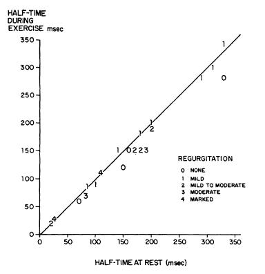
O tempo de meia-pressão (PHT / MP) mantém-se relativamente constante para uma determinada área valvar e sobre uma grande variedade de fluxo. Para uma área valvar de 1cm², o PHT é de 220 ms3. Portanto a área valvar mitral fornecida pelo computador do aparelho é resultante da divisão de 220 pelo PHT achado.
.
Circulation 1968;38;144-150
.
It is concluded that the half-time of the atrioventricular pressure gradient offers a direct, easily determined means for the evaluation of mitral stenosis, even in the presence of complicating mitral regurgitation, or of a hypermetabolic state such as exercise.
.
Ontem fiz um exame de uma gestante no oitavo mês, com diagnóstico de estenose mitral.
Nessa fase da gravidez, a frequencia cardíca sobe e a hipervolemia adicional complica a avaliação.
.
Deu um PHT com área de 1,5 cm2 com gradiente médio de 12 mmhg.
.
O escore de Block deu 8 e a avaliação ao modo B e M sugeria estenose moderada. Pressão pulmonar de 40 mmhg. Refluxo leve.
.
No artigo de 1968, já mostravam que o PHT é confiável mesmo com aumento da frequência e mesmo com vários graus diferentes de regurgitação (figura).
O que não acontece com o gradiente médio que sobe muito com a frequência cardíaca.
.
Após o parto, deve cair bem o gradiente médio e manter o PHT/área.
.
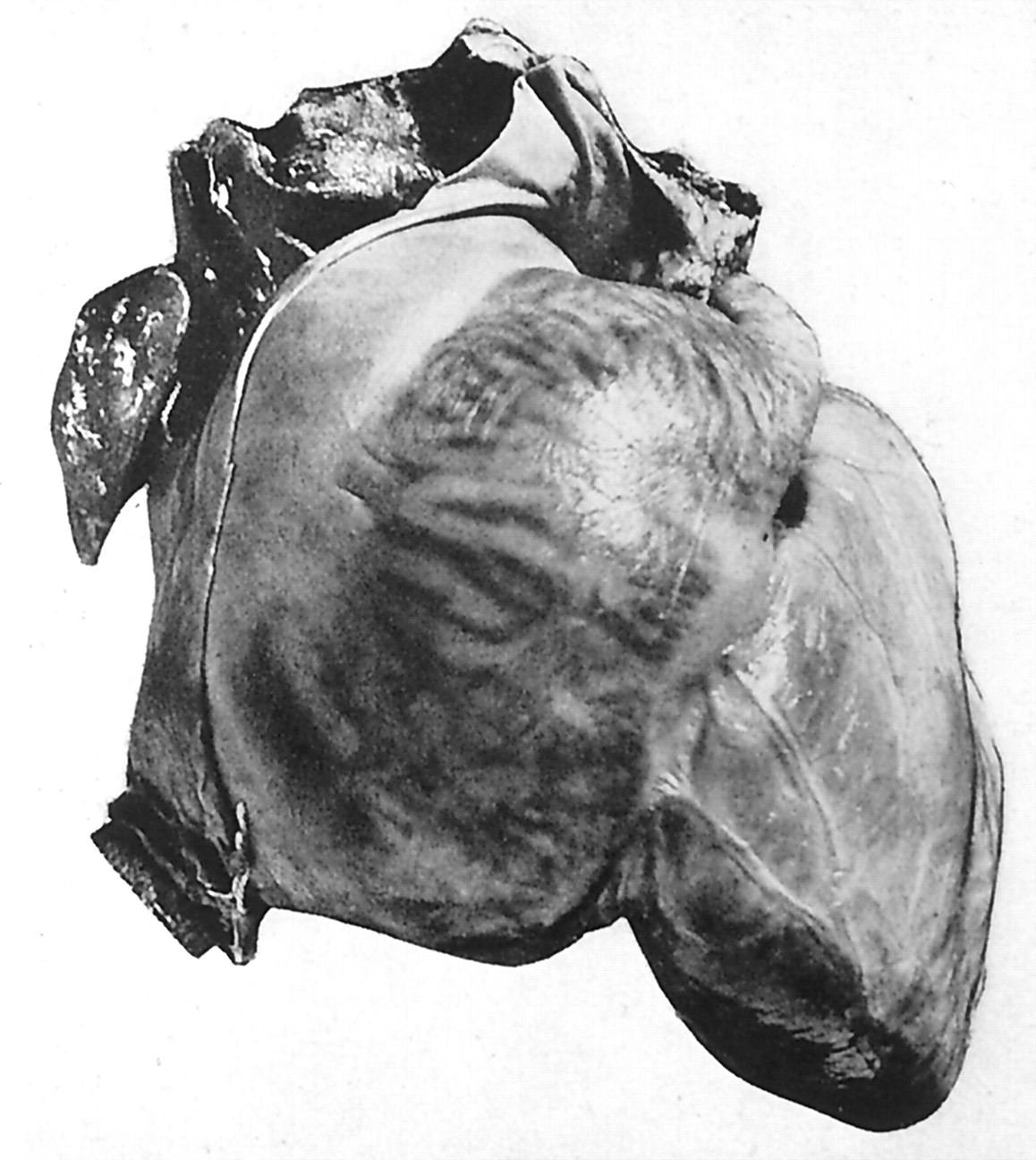
Átrio gigante, só com doença reumática.
.
his photograph was published in the first edition (1931) of Heart Disease. It shows the heart of a patient with a giant left atrium due to predominate mitral regurgitation secondary to rheumatic heart disease. The left atrium held 1760 cc of fluid and the right atrium held 650 cc of fluid. I think this is the same heart that was preserved in a jar in the cardiac museum adjacent to the cardiac conference room in the Massachusetts General Hospital in the late 1940s.
Circulation. 2001;104:2630-2631
.
Autor comenta a ausência de atrios gigantes após o controle dos casos de febre reumática.
Nossos alunos de ecocardiografia contam outra história.
Aqui no país, em estados como Pernambuco, Bahia, Mato Grosso,por exemplo, a febre reumática ainda é frequente, e achados de átrios gigantes são comuns!
.
Trombo no átrio esquerdo.
.
O que determina a formação de um trombo como esse? Estase, lesão endotelial, distúrbio da coagulação?
Aceitamos o achado de trombo facilmente, mas a causa não é determinada de modo exato.
Estase? Ok, mas existe estase venosa quase todos os dias em nossas pernas sedentárias e poucos trombos são formados.
Lesão endotelial no átrio? Possível mas pouco frequente.
Distúrbio da coagulação? Pode ser, mas qual?
E por que permanece fixo no átrio esquerdo, pelo menos por um tempo, não seria mais lógico se soltar?
.
Perguntas que você deve se fazer ao encontrar um trombo.
.