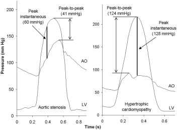
Hipertrofia , estenose e gradientes. Gradiente médio para estenose valvar.

1a

Assessment of Left Ventricular Outflow GradientHypertrophic Cardiomyopathy Versus Aortic Valvular Stenosis

.

http://interventions.onlinejacc.org/article.aspx?articleid=1207395

.

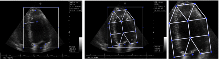
Peak instantaneous gradient was strongly linearly correlated with peak-to-peak gradient in HCM, paralleling the line of identity.

This is opposed to patients with aortic stenosis, where the mean gradient is the standard for both Doppler echocardiography and cardiac catheterization.

.

Nas estenoses valvares aórticas, o gradiente médio é mais confiável.

Cuidado apenas com a frequência cardíaca, que eleva o gradiente médio mesmo sem doença grave.

.

Dica: Quando o gradiente máximo fica muito próximo do gradiente médio, algo está errado.

.

*** Área valvar por continuidade leva a erros absurdos. Residentes medindo área de 0,8 com gradiente máximo de 40 mmHg e médio de 20 mmHg eram frequentes no serviço. O erro estava na área da via de saída, na VTI da via de saída ou nos dois!!! Não acredite nos números sem os outros achados como hipertrofia, dilatação aórtica e gradiente médio.

.

Microbolhas aprovadas com as mesmas restrições nos EUA

1a

.

LINK

.

INDICATIONS

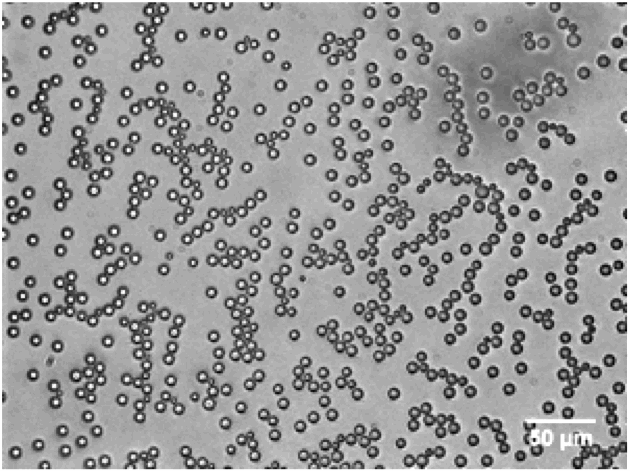
Lumason is an ultrasound contrast agent indicated for use in patients with suboptimal echocardiograms to opacify the left ventricular chamber and to improve the delineation of the left ventricular endocardial border.

.

IMPORTANT SAFETY INFORMATION1

WARNING: SERIOUS CARDIOPULMONARY REACTIONS

Serious cardiopulmonary reactions, including fatalities, have occurred uncommonly during or following the injection of ultrasound contrast agents, including sulfur hexafluoride lipid microspheres  Most serious reactions occur within 30 minutes of administration

  • Assess all patients for the presence of any condition that precludes administration
  • Always have resuscitation equipment and trained personnel readily available

The risk for serious cardiopulmonary reactions may be increased among patients with unstable cardiopulmonary conditions (acute myocardial infarction, acute coronary artery syndromes, worsening or unstable congestive heart failure, or serious ventricular arrhythmias)

.

Integrated backscatter signal (IBS), a nova ressonância de consultório?

1a

.

O ecocardiograma definindo áreas de fibrose!!!

.

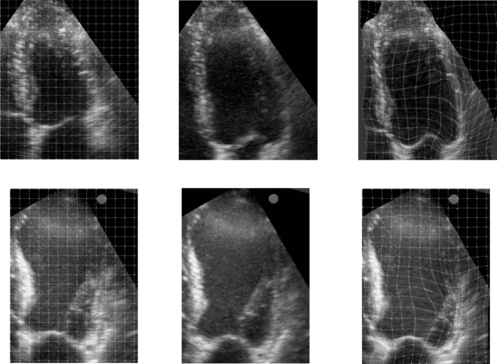
The main finding of this study is represented by the significant difference in echocardiographic indices of fibrosis observed in patients with CRF and in HD treatment in comparison with control subjects with EH. In experimental studies, IBS absolute values have been shown to be reliable indices of LV fibrosis.

.

Myocardial Ultrasound Tissue Characterization in Patients with Chronic Renal Failure

.

http://jasn.asnjournals.org/content/18/6/1953.full

.

Fácil demais de fazer:

.

Images were acquired in parasternal long-axis view (septum and PW were kept almost perpendicular to the ultrasound beam), at a frame rate of 25 Hz, and stored on optical disks. The signal was sampled with the ROI (21 × 21 pixels) placed at the middle portion of the IVS, of the PW, and of the pericardium

.

1a

Pra você que só acredita em colesterol.

1a

.

Metade dos pacientes com doença coronária apresentavam LDL abaixo de 100.

.

1a

.

Fatores de risco tradicionais são bons para grupos, mas falham nos indivíduos.

.

1a

.

Diagnóstico de doença subclínica deve ser feito com exame de baixo custo.

.